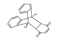
3519-82-2
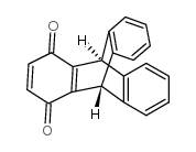
3519-82-2 structure
- Name: INCA-6
- Chemical Name: inca-6
- CAS Number: 3519-82-2
- Molecular Formula: C20H12O2
- Molecular Weight: 284.30800
- Catalog: Research Areas Cardiovascular Disease
- Create Date: 2017-07-04 23:11:41
- Modify Date: 2026-06-30 11:50:11
-
INCA-6 (Triptycene-1,4-quinone) is a cell-permeable NFAT inhibitor. INCA-6 specifically blocks targeting of NFAT(P) substrate to the calcineurin (CN) phosphatase site and is an effective inhibitor of CN-NFAT signaling[1][2][3].
| Name | inca-6 |
|---|---|
| Synonyms |
NFAT Activation Inhibitor III
triptycene quinone Inhibitor of NFAT-Calcineurin Association-6 triptycene-1,4-quinone triptycene-quinine 9,10-dihydro-9,10-o-benzenoanthracene-1,4-dione |
| Description | INCA-6 (Triptycene-1,4-quinone) is a cell-permeable NFAT inhibitor. INCA-6 specifically blocks targeting of NFAT(P) substrate to the calcineurin (CN) phosphatase site and is an effective inhibitor of CN-NFAT signaling[1][2][3]. |
|---|---|
| Related Catalog | |
| In Vitro | INCA-6 (5 μM; for 24-hour) prevents transient outward K+ current (Ito) downregulation in 3-Hz cells[1]. Pre-treatment of BV-2 cells with INCA-6 (10 μM) significantly inhibits ATP-induced CXCL2 expression in BV-2 cells. INCA-6 also inhibits ATP-induced CXCL2 expression in rat primary microglia[2]. INCA-6 (5 μM) reduces SERCA2 transcript levels as well as protein expression, in the absence or in the presence of thapsigargin (TG)[3]. INCA-6 (1.0 and 2.5 μM; 24 hours ) treatment significantly decreases both VEGF and serum-induced human retinal microvascular endothelial cells (HRMEC) proliferation, but does not affect baseline proliferation[4]. Cell Proliferation Assay[4] Cell Line: Human retinal microvascular endothelial cells Concentration: 0.5, 1.0, or 2.5 μM Incubation Time: 24 hours Result: Significantly inhibited VEGF-induced proliferation at 1.0 and 2.5 μM concentrations. |
| In Vivo | INCA-6 (5.0, or 25.0 μM) treatment significantly reduces pathologic neovascularization in oxygen-induced retinopathy (OIR)[4]. Animal Model: Rats bearing OIR model[4] Dosage: 2.5, 5.0, or 25.0 μM Administration: Intravitreal injection on days 14(0) and 14(3) Result: Decreased the severity of OIR in a dose dependent manner. Significant inhibition was seen at 5.0 and 25.0 μM concentrations. |
| References |
| Density | 1.38g/cm3 |
|---|---|
| Boiling Point | 464.2ºC at 760 mmHg |
| Molecular Formula | C20H12O2 |
| Molecular Weight | 284.30800 |
| Flash Point | 172.6ºC |
| Exact Mass | 284.08400 |
| PSA | 34.14000 |
| LogP | 3.28200 |
| Index of Refraction | 1.729 |
| Hazard Codes | Xi |
|---|
|
~84%
3519-82-2 |
| Literature: Zhu, Xiao-Zhang; Chen, Chuan-Feng Journal of Organic Chemistry, 2005 , vol. 70, # 3 p. 917 - 924 |
|
~%
3519-82-2 |
| Literature: Bartlett; Ryan; Cohen Journal of the American Chemical Society, 1942 , vol. 64, p. 2649,2651 |
|
~%
3519-82-2 |
| Literature: Bartlett; Ryan; Cohen Journal of the American Chemical Society, 1942 , vol. 64, p. 2649,2651 |
|
~%
3519-82-2 |
| Literature: Clar Chemische Berichte, 1931 , vol. 64, p. 1676,1679 |
|
~%
3519-82-2 |
| Literature: Clar Chemische Berichte, 1931 , vol. 64, p. 1676,1679 |