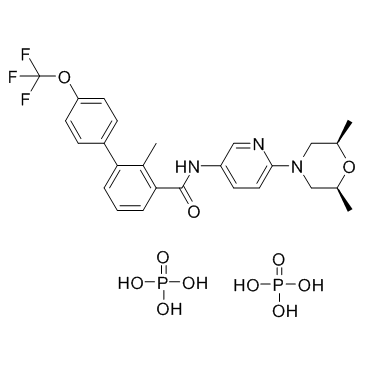
1218778-77-8
| Name | N-[6-[(2S,6R)-2,6-dimethylmorpholin-4-yl]pyridin-3-yl]-2-methyl-3-[4-(trifluoromethoxy)phenyl]benzamide,phosphoric acid |
|---|---|
| Synonyms |
[1,1'-Biphenyl]-3-carboxamide, N-[6-[(2R,6S)-2,6-dimethyl-4-morpholinyl]-3-pyridinyl]-2-methyl-4'-(trifluoromethoxy)-, phosphate (1:2)
LDE225 Diphosphate LDE-225 Diphosphate N-{6-[(2R,6S)-2,6-Dimethyl-4-morpholinyl]-3-pyridinyl}-2-methyl-4'-(trifluoromethoxy)-3-biphenylcarboxamide phosphate (1:2) Sonidegib phosphate LDE 225 Diphosphate UNII:W421AI34UW Odomzo Erismodegib Diphosphate UNII-W421AI34UW NVP-LDE 225 Diphosphate CS-1175 LDE225 (Diphosphate) |
| Description | Erismodegib diphosphate (LDE225 diphosphate) is a potent and selective Smo antagonist with IC50 of 1.3 nM and 2.5 nM for mouse and human Smo in binding assay, respectively. |
|---|---|
| Related Catalog | |
| Target |
IC50: 1.3 nM (mSmo), 2.5 nM (hSmo)[1] |
| In Vitro | The IC50 values for Erismodegib (NVP-LDE225) for the major human CYP450 drug metabolizing enzymes is greater than 10 μM[1]. Erismodegib (LDE225), a small molecule, clinically investigated SMO inhibitor, used alone and in combination with Nilotinib, inhibits the Hh pathway in CD34+ chronic phase (CP)-chronic myeloid leukaemia (CML) cells, reducing the number and self-renewal capacity of CML leukaemia stem cell (LSC). Erismodegib interacts directly with SMO, in a similar fashion to cyclopamine, to reduce expression of downstream Hh signaling targets. Primary CD34+ CP-CML cells are cultured in serum free media (SFM)±Erismodegib for 6, 24 and 72 hours (h). At 72 h, while there is variability between the biological samples, GLI1 is significantly downregulated following exposure to Erismodegib (10 nM; 0.78-fold and 100 nM; 0.73-fold, respectively (p<0.01)[2]. |
| In Vivo | Erismodegib (NVP-LDE225) is a weak base with a measured pKa of 4.2 and exhibits relatively poor aqueous solubility. In the subcutaneous Ptch+/-p53-/- medulloblastoma allograft mouse model, Erismodegib demonstrates dose-related antitumor activity after 10 days of oral administration of a suspension of the diphosphate salt. At a dose of 5 mg/kg/day qd, Erismodegib significantly inhibits tumor growth, corresponding to a T/C value of 33% (p<0.05 as compared to vehicle controls). When dosed at 10 and 20 mg/kg/day qd, Erismodegib affords 51 and 83% regression, respectively[1]. Bone marrow cells and spleen cells from a subset of treated mice are transplanted into secondary recipient mice. Transplantation of either bone marrow (BM) or spleen cells from mice treated with Erismodegib (LDE225)+ Nilotinib results in reduced white cell count (WCC) and reduces leukaemia development in secondary recipients compared to Erismodegib or Nilotinib alone[2]. |
| Cell Assay | CD34+ CP-CML cells are seeded in SFM alone±Erismodegib±Nilotinib and cultured for 24-72 h prior to assessment. Proliferation is measured using colorimetric assessment of BrDU incorporation. Proportion of viable cells versus those in early and late apoptosis is assessed by flow cytometry using annexin V-FITC and 7-amino-actinomycin D (7-AAD, Via-Probe solution). Cell cycle status is assessed using Ki67 (FITC) expression and 7-AAD incorporation. |
| Animal Admin | Mice[2] The transgenic EGFP+/SCLtTA/TRE-BCR-ABL mouse model is used to investigate the effect of Erismodegib treatment on CML LSC in vivo. Scl-tTa-BCR-ABL mice in the FVB/N background are crossed with transgenic GFP-expressing mice. Bone marrow cells are obtained 4 weeks post induction, GFP+ cells are selected by flow cytometry and transplanted by tail vein injection (106 cells/mouse) into wild-type FVB/N recipient mice, irradiated at 900 cGy, generating a large cohort of mice with similar time of onset of leukemia. Blood samples obtained 4 weeks post transplantation confirmed a neutrophilic leukocytosis in recipient mice. Mice are treated with Nilotinib (50 mg/kg by gavage, daily), Erismodegib (80 mg/kg by gavage, daily), Erismodegib+Nilotinib, or with vehicle alone (control). After 3 weeks of treatment, animals are euthanised and marrow content of femurs and tibiae, spleen cells and blood obtained. Total white cell count (WCC), GFP-expressing WCC, myeloid cells, and GFP+ progenitors and stem cells are measured by flow cytometry. Survival is assessed in a subset of mice for 120d post discontinuation of treatment. Spleen and BM cells from a subset of mice in each arm are pooled and 5×10⁶ cells/mouse (8 mice/condition) are transplanted into wild-type FVB/N recipient mice irradiated at 900 cGy. Engraftment is monitored by drawing peripheral blood (PB) every 4 weeks. The percentage of GFP+ cells in PB is analyzed by flow cytometry. |
| References |
| Molecular Formula | C26H32F3N3O11P2 |
|---|---|
| Molecular Weight | 681.489 |
| Exact Mass | 681.146423 |
| PSA | 242.32000 |
| LogP | 4.41330 |
| Storage condition | 2-8℃ |
