| Description |
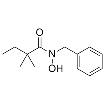
RIPA-56 is a highly potent, selective, and metabolically stable inhibitor of receptor-interacting protein 1 (RIP1) with an IC50 of 13 nM.
|
| Related Catalog |
|
| Target |
IC50: 13 nM (RIP1)[1]
|
| In Vitro |
RIPA-56 has a half-life of 128 min in human liver microsomal stability assays and an EC50 of 28 nM in TSZ-induced HT-29 necrosis assay. RIPA-56 also demonstrates potency in protection of murine L929 cells from TZ-induced necrosis (EC50=27 nM). RIPA-56 shows efficient inhibition of RIP1 kinase activity, with an IC50 of 13 nM and no inhibition of RIP3 kinase activity at a 10 μM concentration. RIPA-56 could form tight hydrophobic interactions with RIP1 through both the phenyl group and the 2,2-dimethylbutyl group, and form two important hydrogen bonds[1].
|
| In Vivo |
In the SIRS mice disease model, RIPA-56 efficiently reduces tumor necrosis factor alpha (TNFα)-induced mortality and multi-organ damage. Compared to known RIP1 inhibitors, RIPA-56 is potent in both human and murine cells, is much more stable in vivo, and is efficacious in animal model studies. RIPA-56 has an impressive PK profile in mice with a 3.1 h half-life, 22% oral bioavailability (P.O.), and 100% bioavailability from intraperitoneal injection (I.P.)[1].
|
| Kinase Assay |
The RIP1 kinase assay is performed in white 384-well plate. RIP1 is first incubated with RIPA-56 or DMSO control for 15 min, then ATP/MBP substrate mixture is added to initiate the reaction. The final concentration of ATP is 50 μM, and MBP 20 μM. After 90 min reaction at room temperature, the ADP-Glo reagent and detection solution are added. The RIP3 kinase assay conditions are almost identical to that of RIP1 assay, except the assay buffer contained 5 mM MgCl2 instead of 20 mM MgCl2 and 12.5 mM MnCl2. The luminescence is measured on PerkinElmer Enspire[1].
|
| Cell Assay |
Cell necrosis assay is performed in 96-well cell culture plate. 3,000 cells are plated in each well and cultured at 37°C overnight. HT-29 cells are treated with 20 ng/mL TNFα/100 nM Smac Mimetics/20 μM z-VAD-FMK and RIPA-56 for 24 h. L929 cells are treated with 20 ng/mL TNFα/20 μM z-VAD-FMK and RIPA-56 for 6 h. The cell survival ratio is determined using the Cell Titer-Glo Luminescent Cell Viability Assay kit[1].
|
| Animal Admin |
Mice: Following intraveneous (IV), intraperitoneal (IP), or oral administration (PO) of RIPA-56 to C57BL/6 mice (n=3), blood is sampled through eye puncture at various time points. Compound concentrations in the plasma samples are analyzed by LCMS/MS. Pharmacokinetic parameters are determined from individual animal data using noncompartmental analysis in phoenix 64[1].
|
| References |
[1]. Ren Y, et al. Discovery of a Highly Potent, Selective, and Metabolically Stable Inhibitor of Receptor-InteractingProtein 1 (RIP1) for the Treatment of Systemic Inflammatory Response Syndrome. J Med Chem. 2017 Feb 9;60(3):972-986.
|
