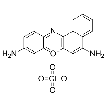
41830-80-2
| Name | cresyl violet perchlorate |
|---|---|
| Synonyms |
CRESYL VIOLET PERCHLORATE
Cresyl Violet 670 CRESYL VIOLET 670 PERCHLORATE) EINECS 255-561-6 Oxazine 9 perchlorate Cresyl Violet perchlorate,laser grade,pure Cresyl Violet perchlorate,laser grade Cresylvioletperchlorate,lasergrade MFCD00012671 Cresyl Violet perchlorate,pure,laser grade |
| Description | Cresyl Violet perchlorate is a red fluorescent stain, which can be used to stain neurons. |
|---|---|
| Related Catalog | |
| In Vitro | The estimated total number of SG neurons is 27,485±3251 and 26,705±1823 in the PV and Cresyl Violet perchlorate (CV) stained sections, respectively. There is no significant difference between them (p=0.552). Therefore, Cresyl Violet perchlorate staining is simpler and more cost effective when estimates neuronal number. Although PV stains spiral ganglion neurons (SGNs) with a greater intensity and provides a functional status, its tedious protocol limits its use for quantification. Total RC volume is estimated using probe and it is found that an average RC volume of 2.162±0.35 mm3 and 1.82±0.33 mm3 in Cresyl Violet perchlorate stained and PV immunostained sections, respectively. Volume of neurons is estimated using nucleator probe and it is 3487.63±951 μm3 and 3740.1±784 μm3 in CV stained and PV immunostained sections, respectively. Similarly, volume of neuronal nucleus is also estimated using nucleator probe and it is found to be 131.68±50 μm3 and 126.51±33 μm3 in CV stained and PV immunostained sections, respectively[1]. |
| Cell Assay | Cochlear sections containing SGNs are placed in 24 wells plates containing PBS (pH 7.4) and stored at 4°C. The sections are then used for Cresyl Violet perchlorate (CV) and immunohistochemical (IHC) staining. Every 7th section is stained with Cresyl Violet perchlorate (1%), dehydrated with ascending grades of alcohol, cleared with xylene, mounted with DPX and observed under microscope. Approximately 12-13 Cresyl Violet perchlorate stained sections from each specimen are used for stereology. None of these cases show any histopathological changes under the light microscope. Estimation of the total volume of the Rosenthal canal (RC), total number of SGNs (optical fractionator probe) and the volume of the soma and their nucleus (nucleator probe) is done with software[1]. |
| References |
| Boiling Point | 482.3ºC at 760mmHg |
|---|---|
| Melting Point | 331 - 333ºC |
| Molecular Formula | C16H12ClN3O5 |
| Molecular Weight | 361.73700 |
| Flash Point | 245.5ºC |
| Exact Mass | 361.04700 |
| PSA | 147.34000 |
| LogP | 3.98470 |
| Index of Refraction | 1.758 |
| Symbol |

GHS07 |
|---|---|
| Signal Word | Warning |
| Hazard Statements | H315-H319-H335 |
| Precautionary Statements | P261-P305 + P351 + P338 |
| Personal Protective Equipment | dust mask type N95 (US);Eyeshields;Gloves |
| Hazard Codes | Xi |
| Risk Phrases | R36/37/38 |
| Safety Phrases | S26-S36 |
| RIDADR | NONH for all modes of transport |
| WGK Germany | 3 |
