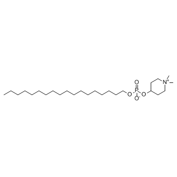
157716-52-4
| Name | perifosine |
|---|---|
| Synonyms |
Piperidinium (4-[[hydroxy(octadecyloxy)phosphinyl]oxy]-1,1-dimethyl
Perifosine (KRX-0401) 4-[[Hydroxy(octadecyloxy)phosphinyl]oxy]-1,1-dimethylpiperidinium Inner Salt Piperidinium, 4-[[hydroxy(octadecyloxy)phosphinyl]oxy]-1,1-dimethyl-, inner salt Perifosine 1,1-Dimethylpiperidinium-4-yl-octadecylphosphat 1,1-Dimethyl-4-piperidiniumyl octadecyl phosphate 1,1-Dimethylpiperidinium-4-yl octadecyl phosphate 1,1-Dimethylpiperidin-1-ium-4-yl octadecyl phosphate (1,1-dimethylpiperidin-1-ium-4-yl) octadecyl phosphate |
| Description | Perifosine is an oral Akt inhibitor. All cells are sensitive to the antiproliferative properties of Perifosine with an IC50 of ~0.6-8.9 μM. |
|---|---|
| Related Catalog | |
| Target |
Akt Autophagy |
| In Vitro | The IC50 for growth of Ntv-a/LacZ cell lines is determined by MTT assay. When the cells are cultured for 48 hours in 10% FCS-supplemented media, the IC50 for cells with constitutively active PDGF, Ras, or Akt signaling is similar and found to be ~45 μM[1].Perifosine, a oral-bioavailable alkylphospholipid (ALK), on the cell cycle kinetics of immortalized keratinocytes (HaCaT) as well as head and neck squamous carcinoma cells. Proliferation is assessed by the incorporation of [3H]thymidine into cellular DNA. Exposure to Perifosine (0.1-30 μM) for 24 h results in a dose-dependent inhibition of [3H]thymidine uptake in all cell lines tested. The IC50s for growth are between 0.6 and 8.9 μM, reaching IC80s of ~10 μM. Perifosine blocks cell cycle progression of head and neck squamous carcinoma cells at G1-S and G2-M by inducing p21WAF1, irrespective of p53 function, and may be exploited clinically because the majority of human malignancies harbor p53 mutations. Perifosine (20 μM) induces both G1-S and G2-M cell cycle arrest, together with p21WAF1 expression in both p53 wild-type and p53-/- clones[2]. |
| In Vivo | Mice are identified with tumors by bioluminescence imaging and either treated them with 100 mg/kg Temozolomide, or 30 mg/kg Perifosine, or a combination with 100 mg/kg Temozolomide and 30 mg/kg Perifosine (Temozolomide+Perifosine) for 3 to 5 days. The mice are sacrificed and tumors analyzed histologically for cell proliferation by Ki-67 immunostaining. Ki-67 staining index is significantly reduced in mice treated with either Temozolomide (Ki-67 staining index=5.5±1.2%, n=4, P=0.0019) or Perifosine (Ki-67 staining index=3.2±1.1%, n=3, P=0.001) compared with Control, demonstrating the inhibitory effect on proliferation. Most importantly, the tumors treated with Temozolomide+Perifosine have the lowest Ki-67 staining index (1.7±1.2%, n=3, P=0.0005). The additional treatment with Perifosine results in a significantly lower proliferation rate than Temozolomide alone (P=0.0087)[1]. Perifosine markedly decreases p-Akt from 10 min to 24 hours and subsequently, moderately decreased p-S6 from 1h to 24 h after injection[3]. |
| Kinase Assay | Exponentially growing cells (HN12, HN30, and HaCaT) are lysed, and 500 μg of total cellular protein are used to immunoprecipitate active cdc2 and cdk2 complexes. After capturing with gammabind G Sepharose and subsequent washes, the active immune complexes are assessed for activity in the presence of increasing concentrations of Perifosine (0.1-30 μM) or flavopiridol (300 nM) in the kinase assay buffer containing [γ-32P]ATP (3000 Ci/mmol) and 0.2 mg/mL histone H1, 25 μM ATP. Reactions are incubated at 37°C for 30 min and terminated by the addition of SDS-gel loading buffer, resolved in SDS-PAGE, and dried gels are subjected to autoradiography and phosphorimaging[2]. |
| Cell Assay | Cell proliferation studies by measuring the uptake of [3H]thymidine is performed. Briefly, HNSCC and HaCaT cells (1-2×10⁴/well) are grown overnight in 24-well plates and exposed to either Perifosine (0.1-30 μM) or PBS (control). After treatment (24-48 h), cells are pulsed with [3H]thymidine (1 μCi/well) for 4-6 h, fixed (5% trichloroacetic acid), and solubilized (0.5 M NaOH) before scintillation counting. Experiments are performed in triplicates[2]. |
| Animal Admin | Mice[1] Drug treatment of tumor-bearing mice. Image-positive Ef-luc Ntv-a mice are treated daily with i.p. administration of buffer alone as a control, or i.p. administration of 100 mg/kg Temozolomide, or oral administration of 30 mg/kg Perifosine, or a combination with Perifosine and Temozolomide for 3 to 5 days. The mean doses of the treatments are: Control, 5 (all five); Temozolomide, 3.75 (three to five); Perifosine, 3.75 (three to four); and Perifosine+Temozolomide, 3 (all three). Control buffer solution consisted of 5% DMSO and 1% Tween 80 in distilled water. Rats[3] To further determine whether the paradoxical effect of rapamycin on S6 phosphorylation is related to upstream signals of Akt-mTOR, rats are treated with Perifosine (20 mg/kg, ip, once), an Akt inhibitor, 30 min before rapamycin administration. Rats are sacrificed 1 h or 6 h after rapamycin injection. |
| References |
| Melting Point | 271-272° (dec) |
|---|---|
| Molecular Formula | C25H52NO4P |
| Molecular Weight | 461.658 |
| Exact Mass | 461.363403 |
| PSA | 68.40000 |
| LogP | 5.60 |
| Appearance | white to beige |
| Storage condition | ?20°C |
| Water Solubility | H2O: soluble10mg/mL, clear |
| Hazard Codes | Xi |
|---|---|
| RIDADR | NONH for all modes of transport |
| HS Code | 2933399090 |
| HS Code | 2933399090 |
|---|---|
| Summary | 2933399090. other compounds containing an unfused pyridine ring (whether or not hydrogenated) in the structure. VAT:17.0%. Tax rebate rate:13.0%. . MFN tariff:6.5%. General tariff:20.0% |


