1342278-01-6
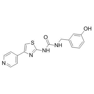
| Name | 1-[(3-hydroxyphenyl)methyl]-3-(4-pyridin-4-yl-1,3-thiazol-2-yl)urea |
|---|---|
| Synonyms |
1-(3-Hydroxybenzyl)-3-[4-(4-pyridinyl)-1,3-thiazol-2-yl]urea
FD5032 RKI-1447 1-(3-hydroxybenzyl)-3-(4-(pyridin-4-yl)thiazol-2-yl)urea 07R |
| Description | RKI-1447 is a potent small molecule inhibitor of ROCK1 and ROCK2 with IC50 values of 14.5 nM and 6.2 nM, respectively. |
|---|---|
| Related Catalog | |
| Target |
ROCK2:6.2 nM (IC50) ROCK1:14.5 nM (IC50) |
| In Vitro | RKI-1447 is a Type I kinase inhibitor that binds the ATP binding site through interactions with the hinge region and the DFG motif. RKI-1447 suppresses phosphorylation of the ROCK substrates mLC-2 and MYPT-1 in human cancer cells, but has no effect on the phosphorylation levels of the AKT, MEK and S6 kinase at concentrations as high as 10 μM. RKI-1447 is also highly selective at inhibiting ROCK-mediated cytoskeleton re-organization. RKI-1447 inhibits migration, invasion and anchorage-independent tumor growth of breast cancer cells[1]. |
| In Vivo | RKI-1447 is highly effective at inhibiting the outgrowth of mammary tumors in a transgenic mouse model. RKI-1447 inhibits mammary tumor growth by 87% and on average the mammary tumors from RKI-1447 treated mice are 7.7 fold smaller compared to those tumors from mice treated with the vehicle control[1]. |
| Kinase Assay | Compounds are tested on three separate days with 8 point dilutions performed in duplicate to determine average IC50 values. The assay conditions are optimized to 15 µL of kinase reaction volume with 5 ng of enzyme in 50 mM HEPES (pH 7.5), 10 mM MgCl2, 1 mM EGTA, and 0.01% Brij-35. The reaction is incubated for 1 h at room temperature in the presence of 1.5 µM of peptide substrate with 12.5 µM of ATP (for ROCK1) or 2 µM of substrate with 50 µM of ATP (for ROCK2). The reaction is then stopped and the ratio of phosphorylated to unphosphorylated peptides is determined by selective cleavage of only the unphosphorylated peptide. The ratio of the signals at 445 nm and 520 nm is measured. IC50 values are determined using fitted curves with GraphPad Prism 5 software[1]. |
| Cell Assay | NIH 3T3 cells are plated at 8000 cells/well in 8-chamber slides in serum free media for 24 hours, and treated with vehicle, 1 μM RKI-1447 or 1 μM RKI-1313 for 1 hour. The cells are then stimulated with complete media, 10 μM LPA, 200 ng/mL bradykinin or 30 ng/mL PDGF for 30 mins. After stimulation, the cells are fixed in 4% paraformaldehyde and stained with Texas-Red Phalloidin. Mounting medium containing DAPI is then added and at least 100 cells per well are observed[1]. |
| Animal Admin | Mice: MMTV/neu transgenic mice are treated i.p. daily for 14 days with either Vehicle [20%-2-hydroxypropyl-beta-cyclodextrin (HPCD)] or 200 mpk RKI-1447 dissolved in freshly prepared HPCD. The percent change in volume is calculated on the basis of the tumor volume on the last day of treatment (Vf) relative to that on the day of initiation of treatment (V0). The average percent change in tumor volume is then calculated for each treatment group[1]. |
| References |
| Density | 1.4±0.1 g/cm3 |
|---|---|
| Molecular Formula | C16H14N4O2S |
| Molecular Weight | 326.373 |
| Exact Mass | 326.083740 |
| PSA | 118.87000 |
| LogP | 1.42 |
| Index of Refraction | 1.701 |
| Storage condition | -20°C |
| Hazard Codes | Xi |
|---|
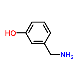
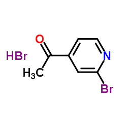
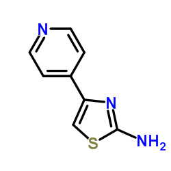
| Precursor 4 | |
|---|---|
| DownStream 0 | |


