1227158-85-1
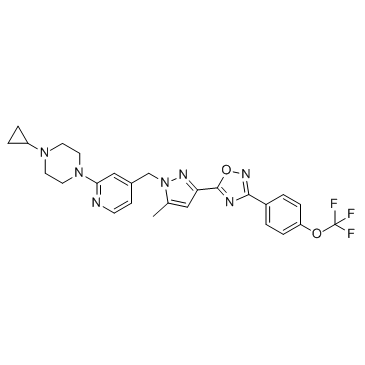
1227158-85-1 structure
- Name: BAY 87-2243
- Chemical Name: 1-cyclopropyl-4-{4-[(5-methyl-3-{3-[4-(trifluoromethoxy)phenyl]-1,2,4-oxadiazol-5-yl}-1H-pyrazol-1-yl)methyl]pyridin-2-yl}piperazine
- CAS Number: 1227158-85-1
- Molecular Formula: C26H26F3N7O2
- Molecular Weight: 525.526
- Catalog: Biochemical Inhibitor Angiogenesis HIF inhibitor
- Create Date: 2018-06-20 14:27:52
- Modify Date: 2026-07-01 01:36:22
-
BAY 87-2243 is a highly potent and selective hypoxia-inducible factor-1 (HIF-1) inhibitor.
| Name | 1-cyclopropyl-4-{4-[(5-methyl-3-{3-[4-(trifluoromethoxy)phenyl]-1,2,4-oxadiazol-5-yl}-1H-pyrazol-1-yl)methyl]pyridin-2-yl}piperazine |
|---|---|
| Synonyms |
bay 87-2243
1-Cyclopropyl-4-{4-[(5-methyl-3-{3-[4-(trifluoromethoxy)phenyl]-1,2,4-oxadiazol-5-yl}-1H-pyrazol-1-yl)methyl]-2-pyridinyl}piperazine |
| Description | BAY 87-2243 is a highly potent and selective hypoxia-inducible factor-1 (HIF-1) inhibitor. |
|---|---|
| Related Catalog | |
| Target |
HIF-1α[1] |
| In Vitro | BAY 87-2243 inhibits luciferase activity with a calculated IC50 value of ~0.7 nM. Hypoxic induction of the HIF target gene CA9 on protein level in HCT116luc cells is inhibited by BAY 87-2243 with an IC50 value of ~2 nM. BAY 87-2243 inhibits mitochondrial oxygen consumption measured by using the oxygen sensitive fluorescence dye LUX-MitoXpress with an IC50 value of ~10 nM[1]. BAY-87-2243 inhibits nuclear HIF-1α protein expression. Administration of BAY-87-2243 for about 18 days significantly reduces HIF-1α protein expression as well as pimonidazole hypoxic fraction (pHF) (mean 2.4% (BAY-87-2243) vs. 17.6% (carrier), p<0.0001), and necrotic fraction (NF) (mean 9% vs. 35.6%, p=0.0002), whereas relative vascular area (RVA) and perfused vessels (PF) remained unchanged[2]. |
| In Vivo | Nude mice are inoculated with H460 cells subcutaneously and after tumors have been established, animals are treated with BAY 87-2243 (0.5, 1, 2, and 4 mg/kg) for 3 weeks by daily oral gavage. BAY 87-2243 reduced tumor weight dose dependently in line with a dose-dependent reduction of the mRNA expression levels of the HIF-1 target genes CA9, ANGPTL4, and EGLN3, whereas the mRNA expression levels of hypoxia-insensitive EGLN2 gene and of HIF-1α itself are not affected by compound treatment in vivo[1]. |
| Cell Assay | Luciferase activity is given in % of DMSO-treated cells. To evaluate the cytotoxicity of BAY 87-2243, 2.000 cells of the respective cell lines are seeded in 96-well plates and cultured in the appropriate growth medium containing 10% FCS. BAY 87-2243 at various concentrations is added at 24 h after seeding for additional 48 h and cell viability is determined using Cell Titer Glow Assay[1]. |
| Animal Admin | Mice[1] Tumor xenograft experiment is carried out on female immune-deficient, athymic NMRI nude mice, aged 7-9 weeks, weighing 20-25 g. The lung carcinoma xenograft mouse model is established by subcutaneous injection into the right flank with 0.1 mL H460 tumor cells (1.5×10⁶) mixed 1:1 with Matrigel. Mice are randomized into control and BAY 87-2243 (0.5, 1, 2, and 4 mg/kg) groups when tumors reach a size of more than 40 mm2. Body weight is monitored as a measure for treatment-related, acute toxicity. Tumor area (measured by caliper) or tumor weight (measured when mice are sacrificed 21 days after cell injection) is calculated by the formula 100-100×(tumor weight/area of treatment group)/(tumor weight/area of vehicle group). Tumor Statistical analysis is performed using the one-way analysis of variance. |
| References |
| Density | 1.5±0.1 g/cm3 |
|---|---|
| Boiling Point | 677.7±65.0 °C at 760 mmHg |
| Molecular Formula | C26H26F3N7O2 |
| Molecular Weight | 525.526 |
| Flash Point | 363.7±34.3 °C |
| Exact Mass | 525.210022 |
| PSA | 85.34000 |
| LogP | 4.20 |
| Vapour Pressure | 0.0±2.1 mmHg at 25°C |
| Index of Refraction | 1.674 |
| Storage condition | -20℃ |