| In Vitro |
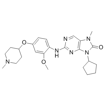
In in vitro kinase assays, AZ3146 inhibits human Mps1Cat with IC50 of ~35 nM. AZ3146 also efficiently inhibits autophosphorylation of full-length Mps1 immunoprecipitated from human cells[1]. TTK specific kinase inhibitor AZ3146 can decrease HCC cell growth. In vitro cell cytotoxicity assays are performed on SMMC-7721 and BEL-7404 cells. IC50s are calculated as being 7.13 μM (BEL-7404) and 28.62 μM (SMMC-7721). Both cells are further treated under the concentration of IC50 for 4 days. Significant inhibitions of cell proliferation are observed[2]. HCT116 cells are cultured for 10 days in 0.8 μM (the GI50) of AZ3146 , then 2 μM AZ3146 for 3 weeks. Sixteen clones are isolated and cell lines generated, named AzR1-16, all of which are resistant to AZ3146-induced cell death in cell viability assays; AzR3 and 4 have a GI50 of approximately 3 μM (4-fold resistance), while the remaining clones have a GI50 of approximately 9 μM (11-fold resistance). When analyzing mitosis by time-lapse microscopy, while 2 μM AZ3146 causes the parental cell line to rapidly exited mitosis in 10 minutes[3].
|
| Kinase Assay |
His-tagged human Mps1Cat encoding amino acids 510-857 is generated. For kinase assays, 500 ng is added to buffer (25 mM Tris-HCl, pH 7.4, 100 mM NaCl, 50 µg/mL BSA, 0.1 mM EGTA, 0.1% β-mercaptoethanol, 10 mM MgCl2, and 0.5 µg/mL myelin basic protein), AZ3146, and 100 µM γ-[32P]ATP (2 µCi/assay). Reactions are incubated at 30°C for 20 min, spotted onto P81 paper, washed in 0.5% phosphoric acid, and immersed in acetone. Phosphate incorporation is determined by scintillation counting. For immunoprecipitation kinase assays, HeLa cells are treated with nocodazole for 14 h, mitotic cells isolated, washed in PBS, and lysed for 30 min in 50 mM Tris-HCl, pH 7.4, 100 mM NaCl, 0.5% NP-40, 5 mM EDTA, 5 mM EGTA, 40 mM β-glycerophosphate, 0.2 mM PMSF, 1 mM DTT, 1 mM sodium orthovanadate, 20 mM sodium fluoride, 1 µM okadaic acid, and complete EDTA-free protease inhibitor cocktail. Full-length Mps1 is immunoprecipitated. Purified complexes are washed with lysis buffer containing 100 mM NaCl and assayed as described for the recombinant protein. To quantify 32P incorporation, reactions are stopped with SDS sample buffer and separated by SDS-PAGE followed by phosphorimaging. The plate is analyzed using a phosphorimager using AIDA software. To assess the specificity of AZ3146, a single-point screen is carried using kinase profiling service. 50 kinases are selected and assayed with 1 µM AZ3146[1].
|
