| Description |
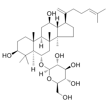
Ginsenoside Rk3 is present in the roots Panax notoginseng herbs. Ginsenoside Rk3 significantly inhibits TNF-α-induced NF-κB transcriptional activity, with an IC50 of 14.24±1.30 μM in HepG2 cells
|
| Related Catalog |
|
| Target |
NF-κB:14.24 μM (IC50, in HepG2 cells)
NF-κB:15.32 μM (IC50, in SK-Hep1 cell)
|
| In Vitro |
Ginsenoside Rk3 exerts the strong activity inhibiting NF-κB in a dose-dependent manner. HepG2 cells are pre-treated with different ginsenosides at concentrations ranging from 0.01 to 10 μM for 1 h, and induced with TNF-α for 20 h. Ginsenoside Rk3 significantly inhibits TNF-α-induced NF-κB transcriptional activity, with an IC50 of 14.24±1.30 μM. Ginsenoside Rk3 significantly inhibits TNF-α-induced NF-κB transcriptional activity, with an IC50 of 15.32±0.29 μM in SK-Hep1 cells, consistent with the data from HepG2 cells. Consistent with the inhibition of NF-κB, Ginsenoside Rk3 inhibits the induction of IL8, CXCL1, iNOS, and ICAM1 mRNA significantly in a dose-dependent manner[1].
|
| In Vivo |
The inhibitory effects of Ginsenoside Rk3 (Rk3) on tumor progression are studied in vivo using a H460 xenograft model in nude mice. Compared with the control group, a significant inhibition of tumor growth (volume) is observed in the Ginsenoside Rk3-treated group. Twenty-one days after treatment initiation, tumor growth is significantly inhibited by approximately 62.99% in the mice receiving 20 mg/kg Ginsenoside Rk3, similar to the inhibitory effect observed in the 20 mg/kg Gefitinib-treated group (57.21%). Compared with the control group, tumor growth is moderately inhibited in the mice receiving 10 and 5 mg/kg Ginsenoside Rk3, with inhibition rates of 32.54% and 11.84%, respectively[2].
|
| Cell Assay |
HepG2 and SK-Hep1 cells are maintained in Dulbecco’s modified Eagle’s medium containing 10% heat-inactivated fetal bovine serum, 100 units/mL Penicillin, and 10 μg/mL Streptomycin, at 37°C and 5% CO2. Cell-Counting Kit (CCK)-8 is used to analyze the effect of compounds (e.g., Ginsenoside Rk3; 0.01, 0.1, 1 and 10 uM) on cell toxicity. Cells are cultured overnight in 96-well plate (~1×10⁴ cells/well). Cell toxicity is assessed after the addition of compounds on dose-dependent manner. After 24 h of treatment, 10 μL of the CCK-8 solution is added to triplicate wells, and incubated for 1 h. Absorbance is measured at 450 nm to determine viable cell numbers in wells[1].
|
| Animal Admin |
Mice[2] Four- to 5-week-old male nude mice are selected and housed under aseptic conditions. The animals are exposed to a phase shift of the light/dark cycle for one week and allowed free access to a normal diet. All animal handling is performed under a laminar flow hood. The H460 xenograft model is established. Cell suspensions at a density of 4-5×10⁶ cells are subcutaneously implanted into the left axilla of each mouse. Tumor engraftment is considered successful when the tumors are clearly visible, which occurs after one to two weeks. The tumor-bearing mice are randomly divided into the following 5 groups (5 mice per group) according to tumor size and body weight: control group (0.9% saline solution), Ginsenoside Rk3-treated group (5/10/20 mg/kg), and Gefitinib-treated group (20 mg/kg). The indicated doses (5-20 mg/kg) of Ginsenoside Rk3 are safe for mice as determined by preliminary acute oral toxicity tests. The dose of Gefitinib is based on the results from another study. Mice are intragastrically treated daily for 21 days. The tumor volumes are estimated. The body weights and tumor volumes of the mice in each group are measured twice per week. After 21 days, all animals are euthanized, and all tumor tissues are removed, weighed, and collected.
|
| References |
[1]. Cho K, et al. Inhibition of TNF-α-Mediated NF-κB Transcriptional Activity by Dammarane-Type Ginsenosidesfrom Steamed Flower Buds of Panax ginseng in HepG2 and SK-Hep1 Cells. Biomol Ther (Seoul). 2014 Jan;22(1):55-61. [2]. Duan Z, et al. Anticancer effects of ginsenoside Rk3 on non-small cell lung cancer cells: in vitro and in vivo. Food Funct. 2017 Oct 18;8(10):3723-3736.
|


