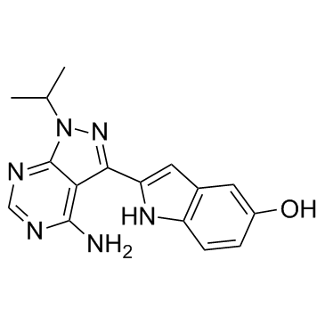
1092351-67-1
| Name | (2E)-2-(4-amino-1-propan-2-yl-2H-pyrazolo[3,4-d]pyrimidin-3-ylidene)indol-5-ol |
|---|---|
| Synonyms |
EX-7207
PP-242 2-[4-Amino-1-(1-methylethyl)-1H-pyrazolo[3,4-d]pyrimidin-3-yl]-1H-indol-5-ol UNII-H5669VNZ7V QCR-154 CS-0247 2-(4-Amino-1-isopropyl-1H-pyrazolo[3,4-d]pyrimidin-3-yl)-1H-indol-5-ol PP 242 2-(4-Amino-1-isopropyl-1H-pyrazolo(3,4-d)pyrimidin-3-yl)-1H-indol-5-ol TORKinib PP242 |
| Description | Torkinib (PP 242) is a selective and ATP-competitive mTOR inhibitor with an IC50 of 8 nM. PP242 inhibits both mTORC1 and mTORC2 with IC50s of 30 nM and 58 nM, respectively. |
|---|---|
| Related Catalog | |
| Target |
mTORC1:30 nM (IC50) mTORC2:58 nM (IC50) mTOR:8 nM (IC50) p110δ:100 nM (IC50) DNA-PK:410 nM (IC50) PDGFR:410 nM (IC50) p110α:2 μM (IC50) p110β:2.2 μM (IC50) p110γ:1.3 μM (IC50) Abl:3.6 μM (IC50) Hck:1.2 μM (IC50) Scr:1.4 μM (IC50) Scr(T338I):5.1 μM (IC50) VEGFR2:1.5 μM (IC50) EGFR:4.4 μM (IC50) EphB4:3.4 μM (IC50) Autophagy Mitophagy |
| In Vitro | Torkinib (PP 242) potently inhibits mTOR (IC50=8 nM) but is much less active against other PI3K family members. Testing of Torkinib (PP 242) against 219 protein kinases reveals remarkable selectivity relative to the protein kinome: at a concentration 100-fold above its IC50 for mTOR, Torkinib (PP 242) inhibits only one kinase by more than 90% (Ret) and only three by more than 75% (PKCα, PKCβII and JAK2V617F)[1]. Torkinib (PP 242) has a dose-dependent effect on proliferation and at higher doses is much more effective than Rapamycin at blocking cell proliferation. The ability of Torkinib (PP 242) to block cell proliferation more efficiently than Rapamycin could be a result of its ability to inhibit mTORC1 and mTORC2, because Rapamycin can only inhibit mTORC1. In SIN1-/- mouse embryonic fibroblasts (MEFs), Rapamycin is also less effective at blocking cell proliferation than Torkinib. That Torkinib (PP 242) and Rapamycin exhibit very different anti-proliferative effects in SIN1-/- MEFs suggests that the two compounds differentially affect mTORC1[2]. |
| In Vivo | In fat and liver, Torkinib (PP 242) is able to completely inhibit the phosphorylation of Akt at S473 and T308, consistent with its effect on these phosphorylation sites observed in cell culture. Surprisingly, Torkinib (PP 242) is only partially able to inhibit the phosphorylation of Akt in skeletal muscle and is more effective at inhibiting the phosphorylation of T308 than S473, despite it's ability to fully inhibit the phosphorylation of 4EBP1 and S6. These results will be confirmed by in vivo dose-response experiments, but, consistent with the partial effect of Torkinib (PP 242) on pAkt in skeletal muscle, a muscle-specific knockout of the integral mTORC2 component rictor resulted in only a partial loss of Akt phosphorylation at S473. These results suggest that a kinase other than mTOR, such as DNA-PK, may contribute to phosphorylation of Akt in muscle[2]. |
| Cell Assay | Wild-type and SIN1-/- MEFs are plated in 96-well plates at approximately 30% confluence and left overnight to adhere. The following day cells are treated with Torkinib (PP 242) (1 nM, 10 nM, 100 nM, 1 μM, and 10 μM), Rapamycin, or vehicle (0.1% DMSO). After 72 h of treatment, 10 μL of 440 μM resazurin sodium salt is added to each well, and after 18 h, the florescence intensity in each well is measured using a top-reading florescent plate reader with excitation at 530 nm and emission at 590 nm[2]. |
| Animal Admin | Mice[2] Six-wk-old male C57BL/6 mice are fasted overnight prior to drug treatment. Torkinib (PP 242) (0.4 mg), Rapamycin (0.1 mg), or vehicle alone is injected IP. After 30 min for the Rapamycin-treated mouse or 10 min for the Torkinib (PP 242) and vehicle-treated mice, 250 mU of insulin in 100 μL of saline is injected IP. 15 min after the insulin injection, the mice are killed by CO2 asphyxiation followed by cervical dislocation. Tissues are harvested and frozen on liquid nitrogen in 200 μL of cap lysis buffer. The frozen tissue is thawed on ice, manually disrupted with a mortar and pestle, and then further processed with a micro tissue-homogenizer. Protein concentration of the cleared lysate is measured by Bradford assay and 5-10 μg of protein is analyzed by Western blot[2]. |
| References |
| Density | 1.6±0.1 g/cm3 |
|---|---|
| Boiling Point | 642.0±50.0 °C at 760 mmHg |
| Molecular Formula | C16H16N6O |
| Molecular Weight | 308.338 |
| Flash Point | 342.1±30.1 °C |
| Exact Mass | 308.138550 |
| PSA | 105.64000 |
| LogP | 1.83 |
| Vapour Pressure | 0.0±2.0 mmHg at 25°C |
| Index of Refraction | 1.800 |
| Hazard Codes | Xi |
|---|


