108409-83-2
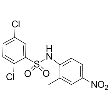
108409-83-2 structure
- Name: FH535
- Chemical Name: 2,5-Dichloro-N-(2-methyl-4-nitrophenyl)benzenesulfonamide
- CAS Number: 108409-83-2
- Molecular Formula: C13H10Cl2N2O4S
- Molecular Weight: 361.201
- Catalog: Biochemical Inhibitor Stem Cells & Wnt Signaling Paths (Stem Cells & Wnt) Wnt/beta-catenin inhibitor
- Create Date: 2018-12-26 19:34:59
- Modify Date: 2026-07-01 17:03:40
-
FH535 is an inhibitor of Wnt/β-catenin and PPAR, with anti-tumor activities.
| Name | 2,5-Dichloro-N-(2-methyl-4-nitrophenyl)benzenesulfonamide |
|---|---|
| Synonyms |
fh535
2,5-Dichloro-N-(2-methyl-4-nitrophenyl)benzenesulfonamide |
| Description | FH535 is an inhibitor of Wnt/β-catenin and PPAR, with anti-tumor activities. |
|---|---|
| Related Catalog | |
| Target |
PPAR Wnt β-catenin |
| In Vitro | FH535 is an inhibitor of Wnt/β-catenin and PPAR. FH535 inhibits PPARγ and PPARδ transactivation in HCT116 cells. FH535 (15 μM) activities depend on functional PPARδ but does not require a cysteine residue in the PPAR ligand-binding domain. FH535 inhibits recruitment of the coactivators GRIP1 and β-catenin to PPARδ and PPARγ. FH535 shows toxic effects on 12 carcinoma cell lines expressing wnt/β-catenin pathway[1]. FH535 (20 μM) suppresses the β-catenin pathway in pancreatic cancer cells, and inhibits pancreatic cancer cell migration. Furthermore, FH535 (20, 40 μM) inhibits pancreatic cancer cell invasion and cell growth[2]. FH535 represses angiogenesis-related genes in pancreatic cancer cells[3]. |
| In Vivo | FH535 (25 mg/kg, i.p.) exhibits an anti-tumor effect on pancreatic cancer xenografts in mice. FH535 also represses angiogenesis in pancreatic cancer xenografts[2]. |
| Cell Assay | Cell growth is evaluated using the MTT assay. Cells (5 × 10⁴/well) are seeded in 24-well tissue culture plates. Blank control is treated with DMSO. After FH535 treatment, MTT is added to each well (final concentration, 0.5 mg/mL), followed by 4-hour incubation at 37°C. The medium is removed, and 800 μL of DMSO is added to each well. The absorbance of the mixture is measured at 490 nm using a microplate enzyme-linked immunosorbent assay reader. The relative cell viability is calculated as follows: relative cell viability = (mean experimental absorbance/mean control absorbance) ×100%[2]. |
| Animal Admin | Four-week-old female BALB/c athymic nude mice receive humane care. PANC-1 cells stably expressing firefly luciferase are injected into the left flanks of the mice in a total volume of 100 μL (0.5 × 10⁷ cells), and the mice are randomly assigned to a DMSO [intraperitoneally injected with 100 μL DMSO/DMEM (1:1)] or FH535 group [intraperitoneally injected with 25 mg/kg FH535 dissolved in 100 μL DMSO/DMEM (1:1)]. Treatment is conducted every 2 days for 20 days; tumor volume is measured with a caliper using the formula: volume = length × width2/2. At the end of the experiment, the mice are anaesthetized and given D-luciferin in PBS. Twenty minutes after the injection, bioluminescence is imaged with a charge-coupled device camera. Then, the tumor tissue is stripped and formalin-fixed, paraffin-embedded, cut into 4-μm sections, and immunohistochemically stained[3]. |
| References |
| Density | 1.6±0.1 g/cm3 |
|---|---|
| Boiling Point | 526.3±60.0 °C at 760 mmHg |
| Melting Point | 141-142℃ |
| Molecular Formula | C13H10Cl2N2O4S |
| Molecular Weight | 361.201 |
| Flash Point | 272.1±32.9 °C |
| Exact Mass | 359.973846 |
| PSA | 100.37000 |
| LogP | 5.12 |
| Vapour Pressure | 0.0±1.4 mmHg at 25°C |
| Index of Refraction | 1.651 |
| Storage condition | 2-8°C |
| Water Solubility | DMSO: soluble20mg/mL |