1373423-53-0
1373423-53-0 structure
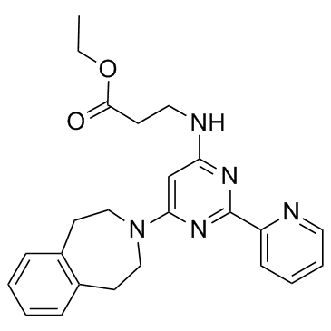
- Name: GSK-J4
- Chemical Name: ethyl 3-[[2-pyridin-2-yl-6-(1,2,4,5-tetrahydro-3-benzazepin-3-yl)pyrimidin-4-yl]amino]propanoate
- CAS Number: 1373423-53-0
- Molecular Formula: C24H27N5O2
- Molecular Weight: 417.504
- Catalog: Biochemical Inhibitor Epigenetics Histone Demethylase Inhibitor
- Create Date: 2017-03-29 10:35:50
- Modify Date: 2026-07-09 01:34:43
-
GSK-J4 is a potent H3K27me3 histone lysine demethylase (KDM) inhibitor, with IC50s of 8.6 μM and 6.6 μM against KDM6B and KDM6A, respectively.
| Name | ethyl 3-[[2-pyridin-2-yl-6-(1,2,4,5-tetrahydro-3-benzazepin-3-yl)pyrimidin-4-yl]amino]propanoate |
|---|---|
| Synonyms |
GSK-J4
Ethyl N-[2-(2-pyridinyl)-6-(1,2,4,5-tetrahydro-3H-3-benzazepin-3-yl)-4-pyrimidinyl]-β-alaninate GSK J4 HCl β-Alanine, N-[2-(2-pyridinyl)-6-(1,2,4,5-tetrahydro-3H-3-benzazepin-3-yl)-4-pyrimidinyl]-, ethyl ester |
| Description | GSK-J4 is a potent H3K27me3 histone lysine demethylase (KDM) inhibitor, with IC50s of 8.6 μM and 6.6 μM against KDM6B and KDM6A, respectively. |
|---|---|
| Related Catalog | |
| Target |
IC50: 8.6 µM (KDM6B), 6.6 µM (KDM6A)[5] |
| In Vitro | GSK-J4 has cellular activity in Flag-JMJD3-transfected HeLa cells, in which GSK-J4 prevents the JMJD3-induced loss of nuclear H3K27me3 immunostaining. Administration of GSK-J4 increases total nuclear H3K27me3 levels in untransfected cells. GSK-J4 significantly reduces the expression of 16 of 34 LPS-driven cytokines, including tumour-necrosis factor-α (TNF-α)[1]. GSK-J4 (10, 25 nM) acts upon DCs promoting the differentiation of Treg cells, improving Treg stability and suppressive capacities, without affecting the differentiation of Th1 and Th17 cells[2]. GSK-J4 inhibits the KDM6 family of H3K27me3 demethylases JMJD3 and UTX. GSK-J4 inhibits JMJD3 expression that is induced by TGF-β1[3]. GSK-J4 inhibits H3K4 demethylation at Xist, Nodal, and HoxC13 in female embryonic stem cells[4]. |
| In Vivo | GSK-J4 (0.5 mg/kg, i.p.) significantly reduces the severity and delays the onset of the disease of the mouse model of experimental autoimmune encephalomyelitis[2]. |
| Animal Admin | Six-to eight-week-old female C57BL/6 WT mice are injected by subcutaneous injection (s.c.) with 50 μg myelin oligodendrocyte glycoprotein 35-55 peptide (pMOG) emulsified in Complete Freund's Adjuvant (CFA) supplemented with heat-inactivated Mycobacterium tuberculosis H37 RA. In addition, mice receive intraperitoneal injection (i.p.) of 500 ng of pertussis toxin on days 0 and 2. Clinical signs are assessed daily according to the following scoring criteria: 0, no detectable signs; 1, flaccid tail; 2, hind limb weakness or abnormal gait; 3, complete hind limb paralysis; 4, paralysis of fore and hind limbs; and 5, moribund or death. A stock solution of GSK-J4 of 42 mg/mL (100 mM) is prepared in dimethyl sulfoxide (DMSO) to preserve stability. Before injection, the stock solution is diluted 1/10 with ethanol (DMSO: ethanol, 1:10 v/v) and brought to a final concentration of 140 μg/mL in PBS. In systemic drug evaluation experiments, each mouse receive daily i.p. injections (from days 0-5) of 100 μL of this solution containing 14.0 μg of the GSK-J4 (equivalent to 0.56 mg/kg of the drug). Control mice receive 100 μL of the vehicle during the same period. In other EAE experiments, 106 bone marrow-derived DCs from WT mice are treated with GSK-J4 or vehicle alone for 16 h, pulsed with 5 μg/mL of pMOG for 4 h and then transferred i.v. into WT C57BL/6 recipient mice 14 and 7 days before EAE induction. In other adoptive transfer EAE experiments, CD4+Foxp3+ Treg cells generated in the presence or absence of 25 nM GSK-J4 are purified by cell sorting and then 0.75×10⁶ transferred i.v. into WT C57BL/6 recipient mice 1 day before EAE induction. |
| References |
[5]. Heinemann B, et al. Inhibition of demethylases by GSK-J1/J4. Nature. 2014 Oct 2;514(7520):E1-2 |
| Density | 1.2±0.1 g/cm3 |
|---|---|
| Boiling Point | 581.2±50.0 °C at 760 mmHg |
| Molecular Formula | C24H27N5O2 |
| Molecular Weight | 417.504 |
| Flash Point | 305.3±30.1 °C |
| Exact Mass | 417.216461 |
| PSA | 80.24000 |
| LogP | 3.75 |
| Appearance | white to beige |
| Vapour Pressure | 0.0±1.6 mmHg at 25°C |
| Index of Refraction | 1.615 |
| Storage condition | 2-8°C |
| Water Solubility | DMSO: soluble20mg/mL, clear |