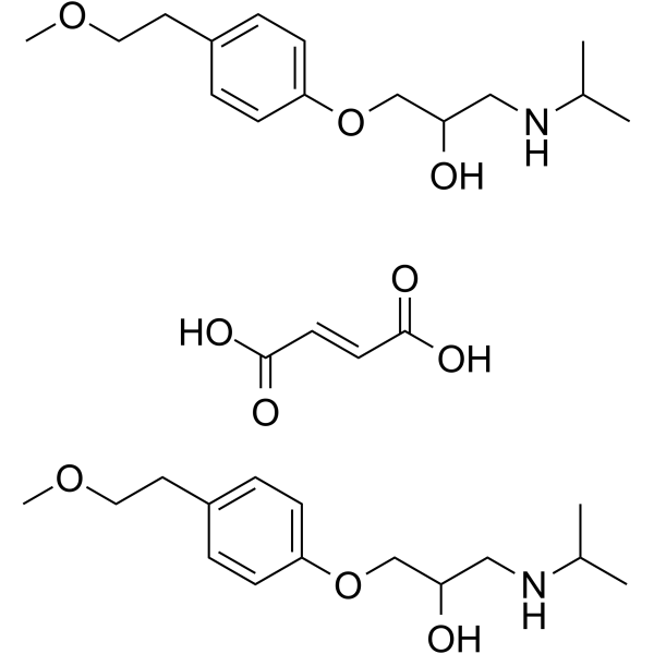
80274-67-5
| Name | Metoprolol fumarate |
|---|---|
| Synonyms |
Metoprolol hemifumarate
1-(Isopropylamino)-3-[4-(2-methoxyethyl)phenoxy]-2-propanol (2E)-2-butenedioate (1:1) 2-Propanol, 1-[4-(2-methoxyethyl)phenoxy]-3-[(1-methylethyl)amino]-, (2E)-2-butenedioate (1:1) (salt) Metoprolol fumarate |
| Description | Metoprolol fumarate (CGP 2175C) is an orally active, selective β1-adrenoceptor antagonist. Metoprolol fumarate shows anti-inflammation, antitumor and anti-angiogenic properties[1][2][3]. |
|---|---|
| Related Catalog | |
| Target |
β1 adrenoceptor |
| In Vitro | Metoprolol (0-1000 μg/mL; 24-72 h) shows cytotoxic effect on U937 and MOLT-4 cells dose and time dependently[3]. Cell Cytotoxicity Assay[3] Cell Line: U937 and MOLT-4 cells Concentration: 1, 10, 50, 100, 500 and 1000 μg/mL Incubation Time: 24, 48 and 72 h Result: Significantly decreased the viability of U937 and MOLT-4 cells at 1000 μg/mL (3740.14µM) concentration after 48 hours incubation time, significantly reduced the viability of U937 cells at ≥500 μg/ml (≥1870.07µM) concentrations after 72 hours incubation time, and significantly decreased the viability of MOLT4 cells at ≥100 μg/ml (≥374.01µM) concentrations after 72 hours incubation. |
| In Vivo | Metoprolol (2.5 mg/kg/h; infusion; 11 weeks) reduces proinflammatory cytokines and atherosclerosis in ApoE−/− Mice[1]. Metoprolol (15 mg/kg/q12h; i.g.; 5 days) shows anti-inflammation and anti-virus effects in murine model with coxsackievirus B3-induced viral myocarditis[2]. Metoprolol (2.5 mg/kg; i.v.; 3 bolus injections) significantly decreased activated caspase-9 protein expression and inhibits myocardial apoptosis in coronary microembolization (CME) rats[4]. Animal Model: Male ApoE−/− mice[1] Dosage: 2.5 mg/kg/h Administration: Via osmotic minipumps, 11 weeks Result: Significantly reduced atherosclerotic plaque area in thoracic aorta, reduced serum TNFα and the chemokine CXCL1 as well as decreasing the macrophage content in the plaques. Animal Model: Balb/c mice, coxsackievirus B3 (CVB3) induced viral myocarditis (VMC) model[2] Dosage: 15 mg/kg/q12h Administration: Oral gavage, 5 consecutive days Result: Reduced pathological scores of VMC induced by CVB3 infection, protected the myocardium against viral damage by reducing serum cTn-I levels. Decreased the levels of myocardial pro-inflammatory cytokines and increase the expression of anti-inflammatory cytokine. Significantly decreased myocardial virus titers. |
| References |
| Molecular Formula | C19H29NO7 |
|---|---|
| Molecular Weight | 383.436 |
| Exact Mass | 383.194397 |
