77603-42-0
77603-42-0 structure
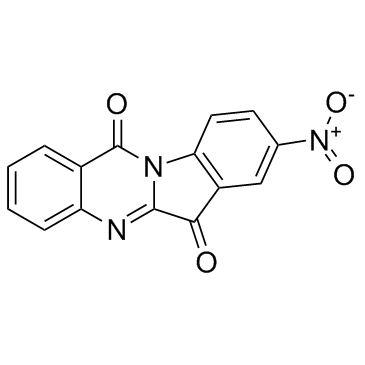
- Name: GNF-PF-3777
- Chemical Name: 8-Nitrotryptanthrin
- CAS Number: 77603-42-0
- Molecular Formula: C15H7N3O4
- Molecular Weight: 293.23
- Catalog: Signaling Pathways Metabolic Enzyme/Protease Indoleamine 2,3-Dioxygenase (IDO)
- Create Date: 2018-06-19 21:31:37
- Modify Date: 2026-07-07 17:17:40
-
8-Nitrotryptanthrin is a potent human indoleamine 2,3-dioxygenase 2 (hIDO2) inhibitor which significantly reduces IDO2 activity with Ki of 0.97 μM.
| Name | 8-Nitrotryptanthrin |
|---|
| Description | 8-Nitrotryptanthrin is a potent human indoleamine 2,3-dioxygenase 2 (hIDO2) inhibitor which significantly reduces IDO2 activity with Ki of 0.97 μM. |
|---|---|
| Related Catalog | |
| Target |
rhIDO2:1.8 μM (IC50) rhIDO2:0.97 μM (Ki) |
| In Vitro | The typtanthrin derivative 8-Nitrotryptanthrin (5i) is found to be a potent hIDO2 inhibitor with superior efficiency far better than that of the most frequently-used inhibitor L-1-MT. The IC50 values show that all nine tryptanthrin compounds display hIDO2 inhibitory activities, especially, the compound 8-Nitrotryptanthrin demonstrates much stronger inhibition (1.87 μM) than both L-1-MT (82.53 μM) and D-1-MT (262.75 μM). 8-Nitrotryptanthrin exhibits significant antitrypanosomal activity with EC50 of 0.82 μM[2]. 8-Nitrotryptanthrin has a microplate Alamar Blue assay (MABA) minimum inhibitory concentration (MIC) value of 0.032 μg/mL. 8-Nitrotryptanthrin also has a LORA MIC value of 2.4 μg/mL, while the majority of analogues lack LORA activity[3]. |
| Kinase Assay | The tryptophan catabolizing activity and enzyme kinetic properties of IDO2 are characterized with mild alterations. Similar to IDO1, IDO2 is also prone to autoxidation. Herein, to maintain the enzyme activity, ascorbic acid (reductant), methylene blue (electron carrier) and catalase (to avoid inhibition of IDO2 by H2O2) are supplemented in the bioassay system. To screen IDO2 inhibitor candidates, each component is dissolved in 50 mM potassium phosphate buffer, pH 7.5. Each assay solution contained 500 μL of reaction mixture comprising 40 mM ascorbic acid, 200 μg/mL catalase, 20 μM methylene blue, substrateL-tryptophan (0-40 mM), the inhibitor candidate (10 μM for preliminary screening and appropriate concentration gradients for Ki and IC50 value determination) and the enzyme at a final concentration of 1500 nM. The assays are conducted at 37°C for 30 min in thermostatically controlled water bath and terminated by addition of 200 μL of trichloroacetic acid (30% w/v). To promote the formation of L-kynurenine, an additional incubation at 65°C for 15 min in water bath is carried out. Each reaction is conducted in triplicate. After centrifuging for 15 min at 13,000× g, 4°C, 100 μL of the supernatant from each tube is transferred into 96-well microplates and mixed with the equal volume of 4-dimethylaminobenzaldehyde (DMAB) in acetic acid (2% w/v). The assay products generated from the dioxygenation of L-Trp catalyzed by IDO2 are confirmed by measuring their absorbance at 492 nm at which wavelength the kynurenine-DMAB complex has an absorption peak. In the assays, the controls are included to eliminate interference, such as any reactions of the inhibitor molecules with the substrate or the substrate with DMAB in the absence of IDO2. For the screening of IDO2 inhibitors, the inhibitory activities of typical competitive inhibitors L-1-MT and D-1-MT are measured under the same conditions as controls[1]. |
| Cell Assay | To study the cellular hIDO2 inhibition of candidate compounds, recombinant plasmid pcDNA3.1(+)-hIDO2 is constructed and transfected into human glioblastoma U87 MG cells which had no IDO1 expression (confirmed by RT-PCR and western blot) therefore eliminated the interference of IDO1. U87 MG cells are cultivated in DMEM containing 50 U/mL penicillin, 50 mg/mL streptomycin, 4500 mg/L glucose, and 10% inactivated FBS at 37°C with 5% CO2 and 95% humidity. When a cell density of 80% confluent monolayer is reached, U87 MG cells are transfected with pcDNA3.1(+)-hIDO2 using the transfection reagent Lipofectamine 2000 according to the manufacturer's instructions. An empty pcDNA3.1(+) expression vector is served as control. After 18 h of incubation, the transfected cells are seeded in 96-well culture plates at a density of 2.5×10⁴ cells/well in a final volume of 200 μL supplemented with 200 μM L-Trp. A serial dilution of the tested compounds is added to the culture medium after an additional 6 h of incubation. The reaction is terminated by addition of 30% (w/v) trichloroacetic acid (10 μL for 140 μL of the reaction mixture) 24 h later. The plates are incubated at 65°C in water bath for 15 min to facilitate the transformation of N-formylkynurenine to L-kynurenine, followed by centrifugation at 13,000× g for 10 min to remove the sediments. 100 μL of the supernatant are then transferred to another 96-well plate and mixed with a same volume of 2% (w/v) 4-dimethylaminobenzaldehyde in acetic acid. The percentages of inhibition of tryptophan degradation or kynurenine production by the compounds are calculated by measuring the absorption at 492 nm using a microplate reader. Cellular IC50s are determined via non-linear regression analysis using GraphPad Prism 5.0[1]. |
| References |
| Molecular Formula | C15H7N3O4 |
|---|---|
| Molecular Weight | 293.23 |
| Water Solubility | DMSO: 6.4 mg/mL (Need warming) |