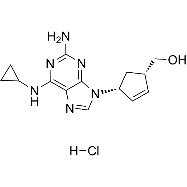
| In Vitro |
Abacavir hydrochloride (15 and 150 μM, 0-120 h) inhibits cell growth, affects cell cycle progression, induces senescence and modulates LINE-1 mRNA expression in prostate cancer cell lines[1]. Abacavir hydrochloride (15 and 150 μM, 18 h) significantly reduces cell migration and inhibits cell invasion[1]. Abacavir hydrochloride induces fat apoptosis[4]. Cell Proliferation Assay[1] Cell Line: PC3, LNCaP and WI-38 Concentration: 15 μM and 150 μM Incubation Time: 0, 24, 48, 72 and 96 hours Result: Showed a dose-dependent growth inhibition on PC3 and LNCaP. Cell Cycle Analysis[1] Cell Line: PC3, LNCaP and WI-38 Concentration: 150 μM Incubation Time: 0, 18, 24, 48, 72, 96 and 120 hours Result: Caused a very high accumulation of cells in S phase in PC3 and LNCaP cells, and G2/M phase increment was observed in PC3 cells. Cell Migration Assay [1] Cell Line: PC3, LNCaP and WI-38 Concentration: 15 and 150 μM Incubation Time: 18 hours Result: Significantly reduced cell migration. Cell Invasion Assay[1] Cell Line: PC3, LNCaP and WI-38 Concentration: 15 and 150 μM Incubation Time: 18 hours Result: Significantly inhibited cell invision.
|
| In Vivo |
Abacavir hydrochloride (100 and 200 mg/kg, p.o.; 4 h) dose-dependently promotes thrombus formation[2]. Abacavir hydrochloride (50 mg/kg/d; i.p.; 14 d) with 0.1 mg/kg/d Decitabine (HY-A0004) enhances survival of high-risk medulloblastoma-bearing mice[3]. Animal Model: Male mice (9-weeks old, 22-30 g) - wild-type (WT) C57BL/6 or homozygous knockout (P2rx7 KO, B6.129P2-P2rx7tm1Gab/J)[2] Dosage: Route 1: 2.5, 5, and 7.5 μg/mL, 100 μL Route 2: 100 and 200 mg/kg Administration: Intrascrotal or oral administration for 4 h Result: Dose-dependently promoted thrombus formation. Animal Model: NSGTM mice, patient-derived xenograft (PDX) cells of non-WNT/non-SHH, Group 3 and of SHH/ TP53-mutated medulloblastoma[3] Dosage: 50 mg/kg/d with 0.1 mg/kg/d Decitabine Administration: Intraperitoneal injection, daily for 14 days Result: Inhibited tumor growth and enhanced mouse survival.
|
