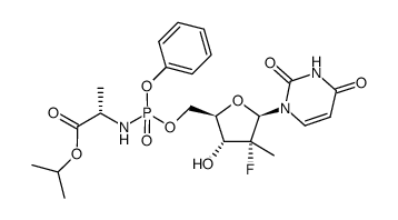
1190307-88-0
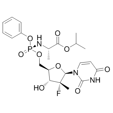
| Name | sofosbuvir |
|---|---|
| Synonyms |
propan-2-yl (2S)-2-[[[(2R,3R,4R,5R)-5-(2,4-dioxopyrimidin-1-yl)-4-fluoro-3-hydroxy-4-methyloxolan-2-yl]methoxy-phenoxyphosphoryl]amino]propanoate
Sovaldi GS-7977 Isopropyl (2S)-2-{[(S)-{[(2R,3R,4R,5R)-5-(2,4-dioxo-3,4-dihydro-1(2H)-pyrimidinyl)-4-fluoro-3-hydroxy-4-methyltetrahydro-2-furanyl]methoxy}(phenoxy)phosphoryl]amino}propanoate (S)-2-{(S)-[(2R,3R,4R,5R)-5-(2,4-dioxo-3,4-dihydro-2H-pyrimidin-1-yl)-4-fluoro-3-hydroxy-4-methyltetrahydrofuran-2-ylmethoxy](phenoxy)phosphorylamino}propionic acid isopropyl ester Sofosbuvir PSI-7977 |
| Description | Sofosbuvir (PSI-7977) is an HCV RNA replication inhibitor with an EC50 of 92 nM. |
|---|---|
| Related Catalog | |
| Target |
EC50: 92±5 nM (HCV)[1] |
| In Vitro | When cathepsin A (CatA) is incubated with PSI-7977 or Sofosbuvir (PSI-7977) for 150 min, ~18-fold more PSI-352707 is formed when Sofosbuvir (PSI-7977) is the substrate compared with PSI-7976. Moreover, the catalytic efficiency for Sofosbuvir (PSI-7977) with CatA is ~30-fold higher than that for PSI-7976[1]. The genotype coverage of Sofosbuvir (PSI-7977) by using GT 1b (Con1)-, 1a (H77)-, and 2a (JFH-1)-derived replicons and GT 1b chimeric replicons containing the NS5B region from the J6 GT 2a isolate and from GT 2b and GT 3a patient isolates is evaluated, Sofosbuvir (PSI-7977) inhibits the replication of these replicons with similar EC50s (between 16 and 48 nM), and is especially active against the chimeric replicon containing the J6 NS5B (EC50=4.7 nM). Sofosbuvir (PSI-7977) inhibits clone A (GT 1b) wild-type and S282T replicons with EC90 values of 0.42 and 7.8 μM, respectively[2]. In the clone A replicon assay, Sofosbuvir (PSI-7977) produces anti-HCV activity with EC90 values 0.42 μM[3]. |
| Cell Assay | Clone A cells are seeded into T75 flasks at about 5×10⁶ cells/flask in Dulbecco's modified Eagle's medium (DMEM) containing 100 IU/mL Penicillin/100 μg/mL streptomycin and 10% fetal bovine serum. Similarly, human primary hepatocytes are seeded in cell plating medium into T75 flasks at about 5×10⁶ cells/flask. After overnight incubation to allow the cells to attach, cells are incubated with 50 μM PSI-7851, PSI-7976, or Sofosbuvir (PSI-7977) in fresh medium for clone A cells or in cell maintenance medium for primary hepatocytes for up to 24 h at 37°C in a 5% CO2 atmosphere. The same procedures are applied when radiolabeled PSI-7851 is used in the study except that 1×10⁶ cells per well are seeded into a 6-well plate, and the cells are incubated with 5 μM [3H]PSI-7851. At selected times, the medium is removed, and the cell layer is washed with cold phosphate-buffered saline (PBS). After trypsinization, cells are counted and centrifuged at 1,200 rpm for 5 min. The cell pellets are suspended in 1 mL of cold 60% methanol and incubated overnight at −20°C. The samples are centrifuged at 14,000 rpm for 5 min, and the supernatants are collected and dried using a SpeedVac concentrator and stored at −20°C until they are analyzed by high performance liquid chromatography (HPLC). Residues are suspended in 100 μL of water, and 50-μL aliquots are injected into HPLC[1]. |
| References |
| Density | 1.4±0.1 g/cm3 |
|---|---|
| Molecular Formula | C22H29FN3O9P |
| Molecular Weight | 529.453 |
| Exact Mass | 529.162537 |
| PSA | 167.99000 |
| LogP | 1.62 |
| Index of Refraction | 1.573 |
| Storage condition | -20°C |
| Hazard Codes | Xi |
|---|---|
| HS Code | 29339900 |
| Precursor 9 | |
|---|---|
| DownStream 0 | |


![L-Alanine, N-[(S)-(4-nitrophenoxy)phenoxyphosphinyl]-, 1-methylethyl ester structure](https://image.chemsrc.com/caspic/082/1256490-31-9.png)
![(S)-2-[(R)-(4-nitro-phenoxy)-phenoxy-phosphorylamino]propionic acid isopropyl ester structure](https://image.chemsrc.com/caspic/061/1256490-49-9.png)
![N-[(S)-(2,3,4,5,6-pentafluorophenoxy)phenoxyphosphinyl]-L-alanine1-Methylethylester structure](https://image.chemsrc.com/caspic/382/1334513-02-8.png)

