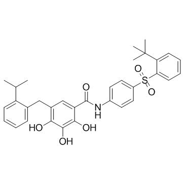
877877-35-5
| Name | N-[4-(2-tert-Butylphenylsulfonyl)phenyl]-2,3,4-trihydroxy-5-(2-isopropylbenzyl)benzamide |
|---|---|
| Synonyms |
N-[4-(2-tert-butylphenyl)sulfonylphenyl]-2,3,4-trihydroxy-5-[(2-propan-2-ylphenyl)methyl]benzamide
2,3,4-Trihydroxy-5-(2-isopropylbenzyl)-N-(4-{[2-(2-methyl-2-propanyl)phenyl]sulfonyl}phenyl)benzamide N-{4-[(2-tert-butylphenyl)sulfonyl]phenyl}-2,3,4-trihydroxy-5-[2-(propan-2-yl)benzyl]benzamide N-[4-[[2-(1,1-Dimethylethyl)phenyl]sulfonyl]phenyl]-2,3,4-trihydroxy-5-[[2-(1-methylethyl)phenyl]methyl]benzamide TW-37 |
| Description | TW-37 is a potent Bcl-2 inhibitor with Ki values of 260, 290 and 1110 nM for Mcl-1, Bcl-2 and Bcl-xL, respectively. |
|---|---|
| Related Catalog | |
| Target |
Mcl-1:260 nM (Ki) Bcl-2:290 nM (Ki) Bcl-xL:1110 nM (Ki) |
| In Vitro | TW-37 (TW37) is a novel nonpeptide small-molecule inhibitor designed using a structure-based design strategy. TW-37 targets the BH3-binding groove in Bcl-2 where proapoptotic Bcl-2 proteins, such as Bak, Bax, and Bid bind. In fluorescence polarization-based binding assays using recombinant Bcl-2 and Bcl-xL proteins, TW-37 binds to Bcl-2 and Bcl-xL with Ki values of 290 and 1110 nM, respectively. TW-37 has an IC50 of 1.8 μM for endothelial cells but shows no cytotoxic effects for fibroblasts at concentrations up to 50 μM. The mechanism of TW-37-induced endothelial cell death is apoptosis, in a process mediated by mitochondrial depolarization and activation of caspase-9 and caspase-3. The effect of TW-37 on endothelial cell apoptosis is not prevented by coexposure to the growth factor milieu secreted by tumor cells. Inhibition of the angiogenic potential of endothelial cells (i.e., migration and capillary sprouting assays) and expression of the angiogenic chemokines CXCL1 and CXCL8 are accomplished at subapoptotic TW-37 concentrations (0.005-0.05 μM)[1]. TW-37 is a potent Bcl-2 and Mcl-1 inhibitor. In fluorescence polarization-based binding assays using recombinant Bcl-2, Bcl-xL, and Mcl-1 proteins, TW-37 binds to Bcl-2, Bcl-xL, and Mcl-1 with Ki values of 290, 1,110 and 260 nM, respectively[2]. |
| In Vivo | A murine model of humanized vasculature is used to investigate the biological effect of TW-37 (TW37) on human microvascular endothelial cell in vivo. Using this model, a significant decrease is observed in total blood vessel number (P<0.05) comparing both 3 and 30 mg/kg TW-37 against vehicle control. In addition to reduction in total number of blood vessels, an unusual number of occluded vessels are occurring in the treated groups. The levels of vessel occlusion are assessed by counting completely blocked vessels and determining their number as a percentage of total vessel number. TW-37 concentration mediates a significant increase in the number of occluded vessels when compared with control[1]. |
| Cell Assay | The sulforhodamine B (SRB) cytotoxicity assay is used. Briefly, optimal cell density for cytotoxicity assay, 2×10⁴ to 3×10⁴ cells per well, is determined by growth curve analysis. HDMECs are seeded at 2.5×10⁴ per well in a 96-well plate and allowed to adhere overnight. Drug or control is diluted in EGM2-MV and layered onto cells, which are allowed to incubate for times as indicated in the figures. Alternatively, HDMECs are coincubated with TW-37 and 0 to 100 ng/mL recombinant human VEGF (rhVEGF)165 or 0 to 100 ng/mL recombinant human CXCL8. Cells are fixed on the plates by addition of cold trichloroacetic acid (10% final concentration) and incubation for 1 hour at 4°C. Cellular protein is stained by addition of 0.4% SRB in 1% acetic acid and incubation at room temperature for 30 minutes. Unbound SRB is removed by washing with 1% acetic acid and the plates are air dried. Bound SRB is resolubilized in 10 mM unbuffered Tris-base and absorbance is determined on a microplate reader at 560 nm. Test results are normalized against initial plating density and drug-free controls. Data are obtained from triplicate wells per condition and are representative of at least three independent experiments[1]. |
| Animal Admin | Mice[1] Porous poly L-lactic acid scaffolds (6×6×1 mm) with an average pore diameter of 180 μm are fabricated. Just before implantation, scaffolds are seeded with 1×10⁶ HDMECs in a 1:1 Matrigel/EGM2-MV mix. Male severe combined immunodeficient (SCID) mice (CB.17.SCID) are anesthetized with ketamine and xylazine, and two scaffolds are implanted s.c. in the dorsal region of each mouse. At 10 days after transplantation, six mice per treatment are treated with 3 mg/kg or 30 mg/kg TW-37 (in vehicle: PBS/Tween 80/ethanol) or vehicle alone i.v. for 5 consecutive days. At the end of the treatment period, mice are euthanized, and the scaffolds are retrieved, fixed overnight in 10% buffered formaldehyde at 4°C, and mounted on glass slides. Immunohistochemistry is done for Factor VIII and microvessels are counted in 6 fields per scaffold and 12 scaffolds per treatment at ×200 magnification. Alternatively, sections are stained with H&E and occluded blood vessels are counted. |
| References |
| Density | 1.3±0.1 g/cm3 |
|---|---|
| Boiling Point | 723.7±60.0 °C at 760 mmHg |
| Molecular Formula | C33H35NO6S |
| Molecular Weight | 573.699 |
| Flash Point | 391.5±32.9 °C |
| Exact Mass | 573.218506 |
| PSA | 132.31000 |
| LogP | 8.74 |
| Vapour Pressure | 0.0±2.4 mmHg at 25°C |
| Index of Refraction | 1.631 |
| Storage condition | Store at -20°C |
