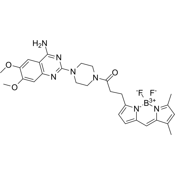
BODIPY FL prazosin
Modify Date: 2024-01-16 12:24:44

BODIPY FL prazosin structure
|
Common Name | BODIPY FL prazosin | ||
|---|---|---|---|---|
| CAS Number | 175799-93-6 | Molecular Weight | 563.41 | |
| Density | N/A | Boiling Point | N/A | |
| Molecular Formula | C28H32BF2N7O3 | Melting Point | N/A | |
| MSDS | N/A | Flash Point | N/A | |
Use of BODIPY FL prazosinBODIPY FL prazosin is a fluorescent α1-adrenergic antagonist with Ki values of 14.5, 43.3 nM for α1a-AR and α1b-AR, respectively. BODIPY FL prazosin also is a fluorescent ligand with the excitation and emission wavelengths are 485 and 535 nm, respectively. BODIPY FL prazosin can be used for study the differences in the subcellular localization of α1-adrenoceptor subtypes[1][2][3]. |
| Name | BODIPY FL prazosin |
|---|
| Description | BODIPY FL prazosin is a fluorescent α1-adrenergic antagonist with Ki values of 14.5, 43.3 nM for α1a-AR and α1b-AR, respectively. BODIPY FL prazosin also is a fluorescent ligand with the excitation and emission wavelengths are 485 and 535 nm, respectively. BODIPY FL prazosin can be used for study the differences in the subcellular localization of α1-adrenoceptor subtypes[1][2][3]. |
|---|---|
| Related Catalog | |
| Target |
α1A-adrenergic receptor:14.5 nM (Ki) α1B-adrenergic receptor:43.3 nM (Ki) |
| In Vitro | BODIPY FL prazosin (10 nM; 30 min at room temperature in 100 μl; COS-7 cells) shows Affinity of various α1-AR ligands with Ki values of 14.5, 43.3 nM for α1a-AR and α1b-AR, respectively[1]. BODIPY FL prazosin (100 nM, 30 min) can be used as molecular probe for the Visualization of the non-adrenoceptor binding site of α1-adrenergic drugs in erythroleukemia cells[3]. |
| References |
| Molecular Formula | C28H32BF2N7O3 |
|---|---|
| Molecular Weight | 563.41 |