| Description |
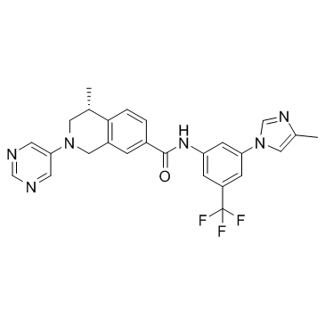
DDR1-IN-3 is a selective Discoidin Domain Receptor 1 (DDR1) inhibitor, with an IC50 value of 9.4 nM.
|
| Related Catalog |
|
| Target |
IC50: 9.4 nM (DDR1)[1].
|
| In Vitro |
DDR1-IN-3 is a promising candidate, with an IC50 value of 9.4 nM against DDR1. DDR1-IN-3 also exhibits reasonable pharmacokinetic (PK) properties, with an oral bioavailability of 66.8% and a T1/2 value of 1.25 h at an oral dose of 20 mg/kg in rats. However, the area under concentration−time curve (AUC) value of DDR1-IN-3 in mice is obviously higher than that in rats, suggesting its good absorption property in mice. The DDR1 inhibition of DDR1-IN-3 is further validated by determining its binding affinity with the DDR1 protein. It is shown that DDR1-IN-3 bounds tightly to DDR1, with a binding constant (Kd) value of 4.7 nM[1].
|
| In Vivo |
DDR1-IN-3 prevents these BLM-induced pathological changes in a dose-dependent manner. These results agree with the expression levels of fibrotic markers in lung tissue lysates, including fibronectin and α-smooth muscle actin (SMA). Further analyses also reveal that the administration of DDR1-IN-3 cause a dose-dependent suppression in the content of hydroxyproline, a unique amino acid found in collagen. The above data collectively indicate the promising therapeutic potential of DDR1-IN-3 against the BLM-induced pulmonary fibrosis[1].
|
| Cell Assay |
Panc-1 cells are plated at low density in media in the presence or absence of controls or the indicated concentration of DDR1-IN-3 (0.016, 0.0625, 0.25, 1 μM). Colony formation is evaluated after 1.5-2 weeks by fixing and staining with crystal violet. The effect of DDR1-IN-3 on cell migration is determined through a ‘scratch’ assay. Panc-1 cells are grown to confluence in a 6 well dish. A scratch is made using a p20 pipette tip and cell migration into the wound is determined at 12, 24, 48, 60, and 72 hrs. The effect of control compounds or DDR1-IN-3 at the indicated concentrations is determined at each time point[1].
|
| Animal Admin |
Mice[1] To induce pulmonary damage, 6- to 8-week-old sex- and age-matched wild type or slie mice (at least five animals per group) are intranasally dropped with bleomycin at 5mg/kg BW. The inhibitors (e.g., DDR1-IN-3) are dissolved in water at a concentration of 5 mg/mL and given to the mice orally by gavage twice a day. Hydroxyproline accounts for 13.4% of the total amino acids of collagen; thus its content can be used to reflect the severity of fibrosis. A commercial hydroxyproline kit is used. Briefly, fresh lung tissues are weighted and hydrolyzed to release hydroxyproline. After a series of chemical reactions, a pink color solution is formed and then subjected to measurement of absorbance at 560 nm. The hydroxyproline content of each sample is calculated by comparing with the standards[1].
|
| References |
[1]. Zhen Wang, et al. Structure-Based Design of Tetrahydroisoquinoline-7-carboxamides as Selective Discoidin Domain Receptor 1 (DDR1) Inhibitors. J Med Chem. 2016 Jun 23; 59(12): 5911–5916.
|