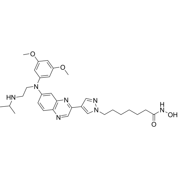
HDAC-IN-50

HDAC-IN-50 structure
|
Common Name | HDAC-IN-50 | ||
|---|---|---|---|---|
| CAS Number | 2653339-26-3 | Molecular Weight | 575.70 | |
| Density | N/A | Boiling Point | N/A | |
| Molecular Formula | C31H41N7O4 | Melting Point | N/A | |
| MSDS | N/A | Flash Point | N/A | |
Use of HDAC-IN-50HDAC-IN-50 is a potent and orally active Apoptosis<0/b> and Apoptosis<1/b> dual inhibitor with IC50 values of 0.18, 1.2, 0.46, 1.4, 1.3, 1.6, 2.6, 13 nM for FGFR1, FGFR2, FGFR3, FGFR4, HDAC1, HDAC2, HDAC6, HDAC8, respectively. HDAC-IN-50 induces Apoptosis and cell cycle arrest at G0/G1 phase. HDAC-IN-50 decreases the expression of pFGFR1,>Apoptosis<2 pSTAT3. HDAC-IN-50 shows anti-tumor activity[1]. |
| Name | HDAC-IN-50 |
|---|
| Description | HDAC-IN-50 is a potent and orally active Apoptosis<0/b> and Apoptosis<1/b> dual inhibitor with IC50 values of 0.18, 1.2, 0.46, 1.4, 1.3, 1.6, 2.6, 13 nM for FGFR1, FGFR2, FGFR3, FGFR4, HDAC1, HDAC2, HDAC6, HDAC8, respectively. HDAC-IN-50 induces Apoptosis and cell cycle arrest at G0/G1 phase. HDAC-IN-50 decreases the expression of pFGFR1,>Apoptosis<2 pSTAT3. HDAC-IN-50 shows anti-tumor activity[1]. |
|---|---|
| Related Catalog | |
| Target |
FGFR1:0.18 nM (IC50) FGFR2:1.2 nM (IC50) FGFR3:0.46 nM (IC50) FGFR4:1.4 nM (IC50) HDAC1:1.3 nM (IC50) HDAC2:1.6 nM (IC50) HDAC6:2.6 nM (IC50) HDAC8:13 nM (IC50) |
| In Vitro | HDAC-IN-50 (compound 10e) (0.1, 1, 10, 100 nM; 12-84 h) induces apoptosis and cell cycle arrest at G0/G1 phase in a time and dose-dependent manner[1]. HDAC-IN-50 (0, 1.25, 2.5, 5 µM for HCT116 cells, 0, 1, 10, 100 nM for SNU-16 cells; 36 h) decreases the expression of pFGFR1, pERK, pSTAT3 in a dose-dependent manner[1]. Cell Proliferation Assay[1] Cell Line: HCT116, SNU-16, KATO III, A2780, K562, Jurkat cells Concentration: 0-30 µM Incubation Time: 72 h Result: Showed antiproliferative activities with IC50s of 0.82, 0.0007, 0.0008, 0.04, 2.46, 15.14 µM for HCT116, SNU-16, KATO III, A2780, K562, Jurkat cells, respectively. Cell Cycle Analysis[1] Cell Line: SNU-16 cells Concentration: 0.1, 1, 10, 100 nM Incubation Time: 12, 24, 36 h Result: Induced cell cycle arrest at G0/G1 phase in a time and dose-dependent manner. Apoptosis Analysis[1] Cell Line: SNU-16 cells Concentration: 0.1, 1, 10, 100 nM Incubation Time: 36, 48, 60, 72, 84 h Result: Induced apoptosis with the apoptotic rate increased 30.8% and 49.6% at 10, 100 nM, respectively. Western Blot Analysis[1] Cell Line: HCT116, SNU-16 cells Concentration: 0, 1.25, 2.5, 5 µM for HCT116 cells, 0, 1, 10, 100 nM for SNU-16 cells Incubation Time: 36 h Result: Reduced the expression of pFGFR1, pERK, pSTAT3 in a dose-dependent manner. |
| In Vivo | HDAC-IN-50 (15, 30 mg/kg; p.o.; daily for 18 days) shows anti-tumor activity in mouse[1]. Pharmacokinetic Parameters of HDAC-IN-50 in female Sprague–Dawley (SD) rats[1]. dose (mg/kg) administration route T1/2 (h) Tmax (h) Cmax (ng/mL) AUC0-∞ (h·ng/mL) CL (mL/min/kg) Vss (mL/kg) F % 2 IV 0.98± 0.12 0.08 1116.63 ± 320.45 424.88 ± 89.56 80.64 ± 15.59 2788.87 ± 765.11 5 IP 1.83 ± 0.06 2 101.57 ± 23.05 491.25 ± 84.18 43.83 30 PO 0.77 ± 0.04 4 442.53 ± 46.33 1557.12 ± 355.61 24.83 Female Sprague-Dawley (SD) rats, 5 mg/kg iv; 5 mg/kg ip; 30 mg/kg p.o.[1] Animal Model: BALB/c nude mice (HCT116 xenograft model)[1] Dosage: 15, 30 mg/kg Administration: P.o.; daily for 18 days Result: Inhibited the tumor growth and downregulated the expression of pSTAT3, pFGFR1, increased the expression of Ac-H3. |
| References |
| Molecular Formula | C31H41N7O4 |
|---|---|
| Molecular Weight | 575.70 |