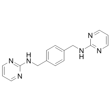
| Description |
MSX-122 is a partial antagonist of CXCR4, inhibiting CXCR4/CXCL12 actions, with an IC50 of ∼10 nM; MSX-122 has anti-inflammatory and anti-metastatic activity.
|
| Related Catalog |
|
| Target |
CXCR4/CXCL12:∼10 nM (IC50)
|
| In Vitro |
MSX-122 is a partial antagonist of CXCR4, inhibiting CXCR4/CXCL12 actions, with an IC50 of ∼10 nM. MSX-122 shows no inhibition on cAMP reduction mediated by their corresponding ligands CCR3/CCL5 and CCR5/CCL5. MSX-122 (100 nM) potently blocks invasion of 78% MDA-MB-231 cells. However, MSX-122 does not suppress T-tropic HIV infection and is inactive in calcium flux assay[1].
|
| In Vivo |
MSX-122 (10 mg/kg, i.p.) blocks inflammation induced by carrageenan and lung fibrosis induced by bleomycin in mice. MSX-122 (4 mg/kg, i.p., daily) blocks metastasis in an experimental animal model of breast cancer metastasis. Furthermore, MSX-122 (10 mg/kg i.p., daily) significantly decreases the numbers of hepatic micrometastases[1].
|
| Animal Admin |
Mice[1] Six- to eight-week-old female nude mice are given injections of 1.5×10⁶ MDA-MB-231 breast cancer cells mixed with the compound (1 µM, less than 5 min preincubation) through the tail vein (10/group). From the following day, mice in the treated group are given 4 mg/kg MSX-122ms (salt form) daily by i.p. injection. The animals are sacrificed 35 days after the tumor cell injection. Whole lung tissues are harvested and sectioned for real-time RT-PCR for human CXCR4 and H&E histostaining to evaluate the metastatic tumor area in five fields per section microscopically. These experiments are repeated once more to confirm the results. For the head and neck cancer animal model, metastatic subclones of 686LN-Ms cells are injected in the same way as MDA-MB-231 cells. [18F]FDG-PET is performed. For the uveal melanoma micrometastasis mouse model, on day 0, each mouse is inoculated with 1×10⁶ wild-type OMM2.3 cells expressing HGF/TGF-β/CXCR4/MMP2 into the posterior chamber of right eye. On day 3, mice are treated with 10 mg/kg MSX-122 in 0.1 mL volume of 45% CD daily by i.p. injection, whereas the control mice are injected with 0.1 mL 45% CD only. On day 7, eyes with tumor are enucleated. The growth of tumor is checked by histological methods. On day 28, hepatic tissues are collected and fixed in 10% formalin, processed, H&E stained, and the number of hepatic micrometastases is counted under microscope. Six sections through the center of the liver are microscopically examined for the presence of micrometastases (<100 µm diameter) and the average number of micrometastases per section is determined. Ten mice per group are used. A table summarizing animal experiments for three metastasis models can be found in the Data S3[1].
|
| References |
[1]. Liang Z, et al. Development of a unique small molecule modulator of CXCR4. PLoS One. 2012;7(4):e34038.
|
