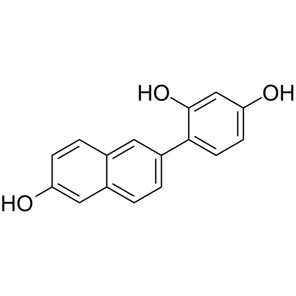
| In Vitro |
HS-1793 (0-100 µM, 24 h) suppresses proliferation of MCF-7, MDA-MB-231 and HCT116 cells[1][2]. HS-1793 (0-50 µM, 4 h) inhibits hypoxia-induced HIF-1α protein in MCF-7 and MDA-MB-231 cells unrelated to cell death, downregulates hypoxia-induced VEGF expression, and suppresses hypoxia-induced mRNA expression of VEGF at the transcriptional level[1]. HS-1793 (0-100 µM, 24 h) induces apoptosis, promotes G2/M cell cycle arrest, and inhibits Akt and ERK phosphorylation in HCT116 cells[2]. Cell Proliferation Assay[1] Cell Line: MCF-7, MDA-MB-231 and MCF-10A Concentration: 0-100 μM Incubation Time: 24 h Result: Showed antiproliferation activity with IC50 values of 26.3±3.2, 48.2±4.2 and >100 μM against MCF-7, MDA-MB-231 and MCF-10A, respectively. Western Blot Analysis[1] Cell Line: MCF-7, MDA-MB-231 Concentration: 12.5, 25 and 50 μM Incubation Time: 4 h Result: Downregulated HIF-1α expression in a concentration-dependent manner in both cell lines. RT-PCR[1] Cell Line: MCF-7, MDA-MB-231 Concentration: 12.5, 25 and 50 μM Incubation Time: 4 h Result: Downregulated the expression of VEGF mRNA, with the more marked results observed in MDA-MB-231 cells. Cell Proliferation Assay[2] Cell Line: HCT116 Concentration: 12.5, 25, 50 and 100 µM Incubation Time: 1, 2 and 4 days Result: Significantly reduced the cell viability concentration- and time-dependently. Significantly suppressed proliferation of colon cancer cell line HCT116. Apoptosis Analysis[2] Cell Line: HCT116 Concentration: 12.5, 25, 50 and 100 µM Incubation Time: 24 h Result: Induced cell apoptosis in a dose-dependent manner. Caused chromatin condensation and fragmentation. Western Blot Analysis[2] Cell Line: HCT116 Concentration: 12.5, 25, 50 and 100 µM Incubation Time: 24 h Result: Effectively induced the reduction of pro-caspase-8 and pro-caspase-3 at 100 µM. Activated caspase-8 and caspase-3. Caused the PARP cleavage. Slightly downregulated the level of antiapoptotic protein Bcl-2 at 100 µM. Promoted an increase in the release of cytochrome c from the mitochondria into the cytosol. Decreased the expression of G2/M cell cycle regulatory protein cyclin B1, Cdc2 and Cdc25C. Decreased the level of CDK4 and CDK6. Decreased Akt phosphorylation and reduced total Akt at high-concentration. Decreased the phosphorylation of ERK1/2 without affecting the protein level. Cell Cycle Analysis[2] Cell Line: HCT116 Concentration: 12.5, 25 and 50 µM Incubation Time: 24 h Result: Induced the accumulation of cells in the G2/M phase in a concentration-dependent manner.
|
