362-07-2
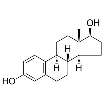
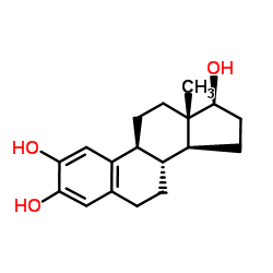
362-07-2 structure
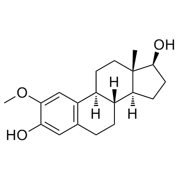
- Name: 2-Methoxyestradiol
- Chemical Name: 2-methoxy-17β-estradiol
- CAS Number: 362-07-2
- Molecular Formula: C19H26O3
- Molecular Weight: 302.408
- Catalog: Biochemical Inhibitor Angiogenesis HIF inhibitor
- Create Date: 2018-07-05 18:12:32
- Modify Date: 2026-07-01 14:34:35
-
2-Methoxyestradiol is an angiogenesis inhibitor and apoptosis inducer with potent antineoplastic activity. 2-Methoxyestradiol also destablize microtubules.
| Name | 2-methoxy-17β-estradiol |
|---|---|
| Synonyms |
(17β)-2-Methoxyestra-1,3,5(10)-triene-3,17-diol
2-methoxy-17beta-estradiol 2-Methoxy 17β-Estradiol 2-Methoxyestra-1,3,5(10)-triene-3,17β-diol 2-Methoxyestradiol 2-Methoxy-b-estradiol 2-Methoxy-β-estradiol MFCD00010489 2-methoxy-17β-estradiol (17b)-2-Methoxyestra-1(10),2,4-triene-3,17-diol |
| Description | 2-Methoxyestradiol is an angiogenesis inhibitor and apoptosis inducer with potent antineoplastic activity. 2-Methoxyestradiol also destablize microtubules. |
|---|---|
| Related Catalog | |
| Target |
Human Endogenous Metabolite |
| In Vitro | 2-Methoxyestradiol (5-100 μM) inhibits assembly of purified tubulin in a concentration-dependent manner, with maximal inhibition (60%) at 200 μM 2-Methoxyestradiol (2ME2). However, with microtubule-associated protein-containing microtubules, significantly higher 2-Methoxyestradiol concentrations are required to depolymerize microtubules, and polymer mass is reduced by only 13% at 500 μM 2-Methoxyestradiol. 4 μM 2-Methoxyestradiol reduces the mean growth rate by 17% and dynamicity by 27%. In living interphase MCF7 cells at the IC50 for mitotic arrest (1.2 μM), 2-Methoxyestradiol significantly suppresses the mean microtubule growth rate, duration and length, and the overall dynamicity, consistent with its effects in vitro, and without any observable depolymerization of microtubules. 2-Methoxyestradiol induces G2-M arrest and apoptosis in many actively dividing cell types while sparing quiescent cells. 2-Methoxyestradiol binds to tubulin at or near the colchicine site, it inhibits microtubule assembly, and high concentrations have been shown to depolymerize microtubules in cells. 2-Methoxyestradiol induces G2-M arrest and apoptosis in many actively dividing also blocks mitosis and inhibits endothelial cell migration[1]. 2-Methoxyestradiol (2-ME) decreases the HIF-1α and HIF-2α nuclear staining in cells cultured under hypoxia. The HIF-1α and HIF-2α mRNA levels are significantly lower when cells are exposed to 2-Methoxyestradiol under normoxia and hypoxia. 2-Methoxyestradiol is an anti-angiogenic, anti-proliferative and pro-apoptotic agent that suppresses HIF-1α protein levels and its transcriptional activity. A significant decrease in the growth rate is found in the 10 µM 2-Methoxyestradiol-treated A549 cells in comparison with the DMSO-treated cells (66.2±7.2 and 101.2±2.3%, respectively; p=0.04) at 96 h. 2-Methoxyestradiol at a concentration of 10 µM is used for the apoptosis and HIF-1α and HIF-2α expression assays, due to the significance found for this concentration when cells are incubated under normoxic conditions at 72 h. A significant increase in apoptosis is observed in cells treated with 10 µM 2-Methoxyestradiol in a normoxic condition in comparison with cells under lower O2 concentration (5.8±0.2%; p=0.003)[2]. |
| In Vivo | To investigate the effect of 2ME2 on uveitis development, C57BL/6 mice are randomly assigned into two groups and immunized with IRBP peptide. 2ME2 group starts 2-Methoxyestradiol (15 mg/kg) intraperitoneally from day 0 to day 13 while control group is given with vehicle. The disease score of 2-Methoxyestradiol (2ME2) group is 0.30±0.30, significantly lower than that of control group 2.09±0.28 (p<0.05), each group containing 5 mice[3]. Treatment with 2-Methoxyestradiol (60-600 mg/kg/d) results in a dose-dependent inhibition of tumor growth. The percentage of cells with strong pimonidazole-positive staining (+++) is significantly decreased in the 2-Methoxyestradiol-treated group (36.0% for 60 mg/kg/d and 0% for 200 and 600 mg/kg/d) compare with the vehicle-treated group (86.5%). This may be attributed to the dramatic inhibition of tumor growth in a dose-dependent manner following 2-Methoxyestradiol treatment[4]. |
| Kinase Assay | Microtubule protein (2.75 mg/mL) is assembled to steady-state [in 100 mM PIPES containing 1 mM EGTA and 1 mM MgSO4 (PEM100) and 1 mM GTP, 35°C for 45 minutes] containing 2-Methoxyestradiol (final drug concentrations of 1-500 μM). Final DMSO and ethanol concentrations are adjusted to 1% and 5%, respectively. Concentrations of 2-Methoxyestradiol ≤ 5 μM have no effect on microtubule polymer mass, and thus 20 to 500 μM 2-Methoxyestradiol is used for most of the experiments. Incubation with 2-Methoxyestradiol is carried out for 30 minutes, at which time microtubule depolymerization is maximal, and microtubules are centrifuged at 35°C for 30 minutes and the supernatant is removed from the pellets. Microtubule pellets are solubilized overnight in 0.2 M NaOH and the protein concentrations of supernatants and pellets are determined[1]. |
| Cell Assay | MCF7 breast carcinoma cells stably transfected with green fluorescent protein (GFP)-α-tubulin are cultured in DMEM supplemented with nonessential amino acids, 0.1% penicillin/streptomycin, 10% fetal bovine serum, and 0.4 mg/mL G418 at 37°C in 5% CO2. Transfection of MCF7 cells with GFP-α-tubulin is carried out. To evaluate mitotic indices, cells are plated at a concentration of 6×10⁴/2 mL into six-well plates. After 48 hours, cells are incubated in the absence or presence of 2-Methoxyestradiol at concentrations ranging from 100 nM to 30 μM for 20 hours. To collect both floating and attached cells, medium is collected; attached cells are rinsed with Versene (137 mM NaCl, 2.7 mM KCl, 1.5 mM KH2PO4, 8.1 mM Na2HPO4, and 0.5 mM EDTA), detached by trypsinization, and added back to the medium. Cells are collected by centrifugation and fixed with 10% formalin for 30 minutes, permeabilized in ice-cold methanol for 10 minutes, and stained with 4′,6-diamidino-2-phenylindole to visualize nuclei. Results are the mean and SE of seven experiments in each of which 500 cells are counted for each concentration. The mitotic IC50 is the drug concentration that induced one half of the maximal mitotic accumulation[1]. |
| Animal Admin | Mice[3] 6~8-week-old C57BL/6 mice are used. C57BL/6 mice are immunized subcutaneously 0.1 mL at tail and 0.05 mL at both thigh sites with IRBP antigen complex. 500 ng Pertussis toxin is injected concurrently. This day is settled as day 0. Then mice are divided into 4 groups, each group containing 5 mice. 15 mg/kg 2-Methoxyestradiol or vehicle is abdominal injected during 0-13 days, 0-6 days, and 7-13 days. At day 14 eyes or lymphoglandula is collected after euthanasia. Rats[4] Fischer 344 rats (average body weight=150 g, n=6 per group) are treated with an i.p. injection of the vehicle (60, 200, or 600 mg/kg/d of 2-Methoxyestradiol/Panzem) for nine consecutive days beginning on the 8th day after the initial tumor cell injection. The experiment is repeated a second time using three rats per group. |
| References |
| Density | 1.2±0.1 g/cm3 |
|---|---|
| Boiling Point | 464.4±45.0 °C at 760 mmHg |
| Melting Point | 188-190°C |
| Molecular Formula | C19H26O3 |
| Molecular Weight | 302.408 |
| Flash Point | 234.7±28.7 °C |
| Exact Mass | 302.188202 |
| PSA | 49.69000 |
| LogP | 3.84 |
| Vapour Pressure | 0.0±1.2 mmHg at 25°C |
| Index of Refraction | 1.586 |
| Storage condition | −20°C |
| Water Solubility | DMSO: 10 mg/mL |
| Symbol |


GHS06, GHS08, GHS09 |
|---|---|
| Signal Word | Danger |
| Hazard Statements | H301 + H311 + H331-H315-H319-H335-H350-H360-H372-H410 |
| Precautionary Statements | P201-P261-P273-P280-P301 + P310-P305 + P351 + P338 |
| Personal Protective Equipment | Eyeshields;Faceshields;full-face particle respirator type N100 (US);Gloves;respirator cartridge type N100 (US);type P1 (EN143) respirator filter;type P3 (EN 143) respirator cartridges |
| Hazard Codes | T: Toxic; |
| Risk Phrases | R23/24/25 |
| Safety Phrases | S22-S26-S36/37/39-S45-S53 |
| RIDADR | UN 3077 9/PG 3 |
| WGK Germany | 3 |
| RTECS | KG7537500 |
| Precursor 6 | |
|---|---|
| DownStream 2 | |