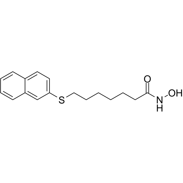
| In Vitro |
HNHA (0-100 μM, 96 h) shows strong inhibition at lower concentrations on cancer cell lines, especially on breast cancer cells, mouse FM3A and human MCF-7[1]. HNHA (15 μM, 24 h) arrests cancer cells at the G1/S phase of the cell cycle, activates p21and rescues strongly protein acetylation[1]. HNHA (15 μM, 12 h) inhibits angiogenic proteins in breast cancer cells, effectively inactivates MMP-2, MMP-9, VEGF and HIF-1α[1]. Cell Proliferation Assay Cell Line: FM3A, C1300, LA-N-1, LA-N-2, LA-N-5, NB16, NB19, NB69, SK-N-SH, MCF-7 and HT-29[1] Concentration: 0-100 μM Incubation Time: 96 h Result: Showed strong inhibition at lower concentrations on all cancer cell lines (FM3A, C1300, LA-N-1, LA-N-2, LA-N-5, NB16, NB19, NB69, SK-N-SH, MCF-7 and HT-29), with IC50 values of 15.70, 55.63, 22.78, 23.18, 26.70, 19.64, 21.26, 22.31, 65.09, 14.33, and 16.98 μM, respectively. Cell Viability Assay Cell Line: FM3A and MCF-7[1] Concentration: 0, 0.1, 1, 5, 10, 15, 20, 25,30 μM Incubation Time: 48 h Result: Showed dose-dependent inhibition of viability in mouse and human breast cancer cells. Cell Cycle Analysis Cell Line: FM3A and MCF-7 cells[1] Concentration: 15 μM Incubation Time: 24 h Result: Arrested FM3A and MCF-7 cells in the G1/S phase. Western Blot Analysis Cell Line: FM3A and MCF-7 cells[1] Concentration: 0, 0.1, 1, 10, and 20 μM (24 h) Incubation Time: 1, 6, 24, 48, and 72 h (15 μM) Result: Activated a cell proliferation arrestor p21, increased histone and non-histone protein acetylation and inhibited FM3A and MCF-7 proliferation in vitro, and was very effective in increasing the acetylation level of histone H3 protein in FM3A and MCF-7. The most effective dose point for acetylation of histone H3 was 10-20 μM. Histone H3 acetylation peaked after 1 h of exposure to the drugs and remained stable for 1-6 h. Western Blot Analysis Cell Line: FM3A and MCF-7 cells[1] Concentration: 15 μM Incubation Time: 12 h Result: Showed a strong induction of TIMP-1 and TIMP-2, and effectively inactivated MMP-2, MMP-9, VEGF and HIF-1α.
|
| In Vivo |
HNHA (20 μM/mouse, IP, once every 2 days for a total of six injections) reduces tumor burden and extends the survival rate, activates TIMP-1, TIMP-2 and p21 and inhibits MMP-2, MMP-9, HIF-1α and VEGF protein expression[1]. Animal Model: C3H/HeJ-FasL mice (FM3A breast cancer cell tumor xenograft, 6 weeks, n = 25/group)[1] Dosage: 20 μM/mouse Administration: IP, once every 2 days for a total of six injections Result: Reduced tumor burden and extended the survival rate. Effectively inhibited cancer development and angiogenesis in vivo. Increased TIMP-1, TIMP-2 and p21, decreased MMP-2, MMP-9, HIF-1α and VEGF protein expression, and reduced the distribution of CD34, HIF-1α and VEGF.
|
