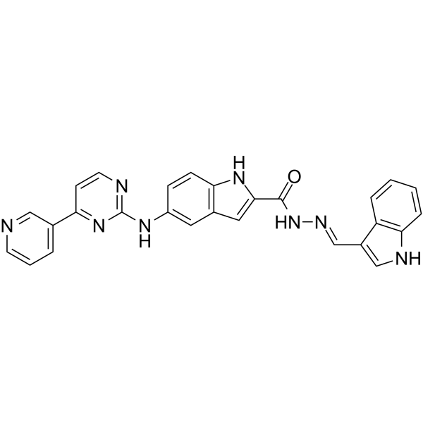
| In Vitro |
CDK9-IN-18 (compound 12i) (0-20 μM, 3 hours; NH2 cells) suppress both HIV-1 transcription and the phosphorylation at Serine 2 of the RNAPII CTD in a dose-dependent manner[1]. CDK9-IN-18 (compound 12i) (0-5.0 μM, 24 hours; human tumor cell lines) exerts antiproliferative effect through the induction of apoptosis in HepG2 cells[1]. Western Blot Analysis[1] Cell Line: NH2 cells Concentration: 0.1, 0.2, 0.5, 1.0 and 2.0 μM Incubation Time: 3 hours Result: P-Ser2 level of RNAPII CTD decreased in a dose-dependent fashion. Cell Cytotoxicity Assay[1] Cell Line: A375 (skin cancer), A549 (lung cancer), HepG2 (liver cancer) and MCF-7 (breast cancer) Concentration: 2.0 μM Incubation Time: 24 hours Result: Inhibited with IC50 values of 0.10, 0.53, 0.07 and 0.10 μM for A375, A549, HepG2 and MCF-7 cells, respectively. Western Blot Analysis[1] Cell Line: A375 (skin cancer), A549 (lung cancer), HepG2 (liver cancer) and MCF-7 (breast cancer) Concentration: 0.1, 0.2, 0.5, 1.0, 2.0 and 5.0 μM Incubation Time: 24 hours Result: The expression level of the specific apoptosisassociated protein (cleaved PARP) increased in a dose-dependent fashion. Cell Cytotoxicity Assay[1] Cell Line: HepG2 cells Concentration: 1.0 and 5.0 μM Incubation Time: 24 hours Result: The percentage of cells in late apoptosis was recorded as 24.2% and 36.3% at 1.0 and 5.0 μM concentrations, respectively.
|
