| Description |
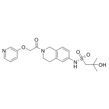
Nampt-IN-1 (LSN3154567) is a potent and selective NAMPT inhibitor. Nampt-IN-1 inhibits purified NAMPT with an IC50 of 3.1 nM.
|
| Related Catalog |
|
| Target |
IC50: 3.1 nM (NAMPT), 0.84 μM (CSF1R)[1]
|
| In Vitro |
To assess its selectivity, specificity, and effects on cellular NAD+ levels, LSN3154567 is characterized in various biochemical and cellular assays. LSN3154567 inhibits purified NAMPT with an IC50 of 3.1 nM. When tested against a panel of human kinases (>100; CEREP Kinase panel), it does not exhibit any significant activity (i.e.: IC50≥1 μM) against the kinases tested except CSF1R (IC50≈0.84 μM). LSN3154567 exhibits a broad spectrum of anticancer activity. To assess its anticancer activity, LSN3154567 is tested against a number of different types of cancer cell lines cultured in the absence or presence of nicotinic acid (NA) (10 μM). LSN3154567 exhibits a potent antiproliferative activity against many cell lines in the absence of NA[1].
|
| In Vivo |
Nampt-IN-1 (LSN3154567) exhibits good physical chemical properties that allow oral dosing. When dosed orally with 2 mg/kg in mice, it has an exposure of 195 nM*hour in the plasma with a peak concentration of 57 nM (at 0.25 hour) and an oral bioavailability of 39%. When dosed intravenously with 2 mg/kg, it has a hepatic clearance of 158.73 mL/min/kg and a volume of distribution at 7.1 L/kg. The half-life of terminal elimination is estimated to be 2.76 hours. LSN3154567 exhibits a dose-dependent inhibition of NAD+ formation with estimated TED50 value of 2.0 mg/kg. To assess whether LSN3154567 causes retinopathy, rats are treated with LSN3154567 at 20, 40, and 80 mg/kg for 4 days. No apparent retinopathy is observed. The hematological toxicities are observed. When LSN3154567 is dosed at 20, 40, and 80 mg/kg, the plasma exposures obtained are 8,974, 18,061, and 38,327 M*h, respectively. Thus, LSN3154567 exhibits exposure multiples of respective 3-, 7-, and 14-fold over the exposure (2,701 M*h) required for robust efficacy (≈103%) without NA coadministration. Dogs are treated with LSN3154567 at 1 and 2.5 mg/kg. At these dose levels, the retinal toxicity is observed. Degeneration of the outer nuclear layer occurred in all four animals, but is less pronounced in the animals treated with 1 mg/kg. At the 1 and 2.5 mg/kg dose levels, the plasma exposures are determined to be 1,483 and 2,468 nM*h, respectively[1].
|
| Cell Assay |
Cell[1]The cell lines used as following: A2780 and KM-12, KMS-11 and MKN-74, OPM-2 and Kelly; and all other cancer cells. The identity of these cell lines is not tested or verified prior to use in this study. Cells are seeded in 96-well plates, cultured overnight, and treated with LSN3154567 (0.03 to 1,000 nM)±NA (10 μM) in duplicate at 37°C in 5% CO2 for 72 hours. Staurosporine (10 μM) is used as positive control. Cell viability is determined by a CytoTox-Glo Cytotoxicity assay kit[1].
|
| Animal Admin |
Mice[1] Male CD-1 mice are dosed with LSN3154567 at 2 mg/kg intravenously in 20% Captisol (w/v), 25 mmol/L NaPO4, pH 2, q.s. formulation or 2 mg/kg orally in the same vehicle in a cross over design with 3-d wash period in between two arms. Blood samples are obtained through retro orbital at 0, 0.08 (IV only), 0.25, 0.75, 2, 4, 8, and 24 hours and frozen until analysis. For exposure analysis, blood samples (≈20 μL) are obtained through tail clipping at 0.5, 1, 2, 4, 8, and 24 hours. The samples are collected into EDTA-coated capillary tubes and spotted onto Whatman DMPK-C DBS collection cards. Rats and Dogs[1] Female rats or male and female dogs are dosed with LSN3154567 orally in the same vehicle. For NA rescue studies, NA is formulated in phosphate buffered saline (pH 7.4). Rats are given LSN3154567 orally once daily at 20, 40, or 80 mg/kg for two cycles of 4 days with 3 days between the first and second cycles. Dogs are also dosed orally at 1 or 2.5 mg/kg (without NA) or at 5 mg/kg (with NA; BID) for 4 days. Exposure is obtained and analyzed as described above.
|
| References |
[1]. Zhao G, et al. Discovery of a Highly Selective NAMPT Inhibitor That Demonstrates Robust Efficacy and Improved Retinal Toxicity with Nicotinic Acid Coadministration. Mol Cancer Ther. 2017 Dec;16(12):2677-2688.
|
